7CL6 | pdb_00007cl6

The crystal structure of KanJ in complex with neamine and N-oxalylglycine


Experimental Data Snapshot

  • Method: X-RAY DIFFRACTION
  • Resolution: 2.44 Å
  • R-Value Free: 
    0.220 (Depositor), 0.222 (DCC) 
  • R-Value Work: 
    0.190 (Depositor), 0.194 (DCC) 
  • R-Value Observed: 
    0.191 (Depositor) 

Starting Model: experimental
View more details

wwPDB Validation 3D Report Full Report

Validation slider image for 7CL6

Ligand Structure Quality Assessment 


This is version 1.3 of the entry. See complete history

Literature

Stepwise Post-glycosylation Modification of Sugar Moieties in Kanamycin Biosynthesis.

Kudo, F.Kitayama, Y.Miyanaga, A.Numakura, M.Eguchi, T.

(2021) Chembiochem 22: 1668-1675

  • DOI: https://doi.org/10.1002/cbic.202000839
  • Primary Citation Related Structures: 
    7CL2, 7CL3, 7CL4, 7CL5, 7CL6

  • PubMed Abstract: 

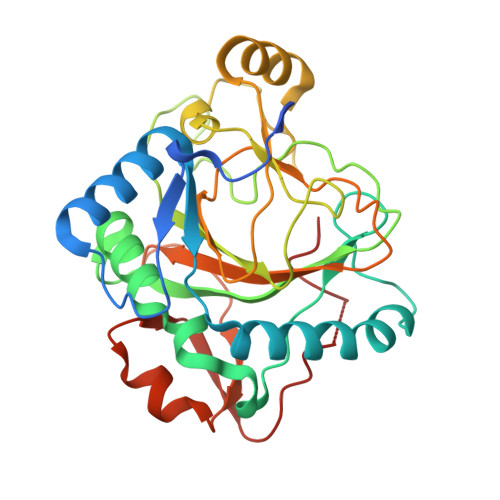
    Kanamycin A is the major 2-deoxystreptamine (2DOS)-containing aminoglycoside antibiotic produced by Streptomyces kanamyceticus. The 2DOS moiety is linked with 6-amino-6-deoxy-d-glucose (6ADG) at O-4 and 3-amino-3-deoxy-d-glucose at O-6. Because the 6ADG moiety is derived from d-glucosamine (GlcN), deamination at C-2 and introduction of C-6-NH 2 are required in the biosynthesis. A dehydrogenase, KanQ, and an aminotransferase, KanB, are presumed to be responsible for the introduction of C-6-NH 2 , although the substrates have not been identified. Here, we examined the substrate specificity of KanQ to better understand the biosynthetic pathway. It was found that KanQ oxidized kanamycin C more efficiently than the 3''-deamino derivative. Furthermore, the substrate specificity of an oxygenase, KanJ, that is responsible for deamination at C-2 of the GlcN moiety was examined, and the crystal structure of KanJ was determined. It was found that C-6-NH 2 is important for substrate recognition by KanJ. Thus, the modification of the GlcN moiety occurs after pseudo-trisaccharide formation, followed by the introduction of C-6-NH 2 by KanQ/KanB and deamination at C-2 by KanJ.


  • Organizational Affiliation
    • Department of Chemistry, Tokyo Institute of Technology, 2-12-1 O-okayama, Meguro-ku, Tokyo, 152-8551, Japan.

Macromolecule Content 

  • Total Structure Weight: 204.79 kDa 
  • Atom Count: 13,557 
  • Modeled Residue Count: 1,678 
  • Deposited Residue Count: 1,806 
  • Unique protein chains: 1

Macromolecules

Find similar proteins by:|  3D Structure
Entity ID: 1
MoleculeChains  Sequence LengthOrganismDetailsImage
Kanamycin B dioxygenase
A, B, C, D, E
A, B, C, D, E, F
301Streptomyces kanamyceticusMutation(s): 0 
Gene Names: kanJkacB
EC: 1.14.11.37
UniProt
Find proteins for Q6L732 (Streptomyces kanamyceticus)
Explore Q6L732 
Go to UniProtKB:  Q6L732
Entity Groups
Sequence Clusters30% Identity50% Identity70% Identity90% Identity95% Identity100% Identity
UniProt GroupQ6L732
Sequence Annotations
Expand
Reference Sequence

Small Molecules

Ligands 3 Unique
IDChains Name / Formula / InChI Key2D Diagram3D Interactions
XXX
(Subject of Investigation/LOI)

Query on XXX


Download:Ideal Coordinates CCD File
I [auth A]
L [auth B]
O [auth C]
R [auth D]
U [auth E]
I [auth A],
L [auth B],
O [auth C],
R [auth D],
U [auth E],
X [auth F]
(1R,2R,3S,4R,6S)-4,6-diamino-2,3-dihydroxycyclohexyl 2,6-diamino-2,6-dideoxy-alpha-D-glucopyranoside
C12 H26 N4 O6
SYJXFKPQNSDJLI-HKEUSBCWSA-N
OGA
(Subject of Investigation/LOI)

Query on OGA


Download:Ideal Coordinates CCD File
H [auth A]
K [auth B]
N [auth C]
Q [auth D]
T [auth E]
H [auth A],
K [auth B],
N [auth C],
Q [auth D],
T [auth E],
W [auth F]
N-OXALYLGLYCINE
C4 H5 N O5
BIMZLRFONYSTPT-UHFFFAOYSA-N
NI
(Subject of Investigation/LOI)

Query on NI


Download:Ideal Coordinates CCD File
G [auth A]
J [auth B]
M [auth C]
P [auth D]
S [auth E]
G [auth A],
J [auth B],
M [auth C],
P [auth D],
S [auth E],
V [auth F]
NICKEL (II) ION
Ni
VEQPNABPJHWNSG-UHFFFAOYSA-N

Experimental Data & Validation

Experimental Data

  • Method: X-RAY DIFFRACTION
  • Resolution: 2.44 Å
  • R-Value Free:  0.220 (Depositor), 0.222 (DCC) 
  • R-Value Work:  0.190 (Depositor), 0.194 (DCC) 
  • R-Value Observed: 0.191 (Depositor) 
Space Group: P 1 21 1
Unit Cell:
Length ( Å )Angle ( ˚ )
a = 50.34α = 90
b = 185.227β = 94.3
c = 109.493γ = 90
Software Package:
Software NamePurpose
REFMACrefinement
PDB_EXTRACTdata extraction
XDSdata reduction
SCALAdata scaling
MOLREPphasing

Structure Validation

View Full Validation Report


Ligand Structure Quality Assessment 


Entry History 

Deposition Data

Revision History  (Full details and data files)

  • Version 1.0: 2021-01-13
    Type: Initial release
  • Version 1.1: 2021-01-20
    Changes: Database references
  • Version 1.2: 2021-05-12
    Changes: Database references
  • Version 1.3: 2023-11-29
    Changes: Data collection, Database references, Refinement description