7CJ3

Crystal structure of the transmembrane domain of Salpingoeca rosetta rhodopsin phosphodiesterase


Experimental Data Snapshot

  • Method: X-RAY DIFFRACTION
  • Resolution: 2.60 Å
  • R-Value Free: 0.295 
  • R-Value Work: 0.246 
  • R-Value Observed: 0.249 

wwPDB Validation   3D Report Full Report


Ligand Structure Quality Assessment 


This is version 1.2 of the entry. See complete history


Literature

Structural insights into the mechanism of rhodopsin phosphodiesterase.

Ikuta, T.Shihoya, W.Sugiura, M.Yoshida, K.Watari, M.Tokano, T.Yamashita, K.Katayama, K.Tsunoda, S.P.Uchihashi, T.Kandori, H.Nureki, O.

(2020) Nat Commun 11: 5605-5605

  • DOI: https://doi.org/10.1038/s41467-020-19376-7
  • Primary Citation of Related Structures:  
    7CJ3, 7D7P, 7D7Q

  • PubMed Abstract: 

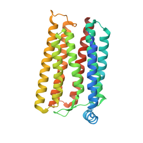
    Rhodopsin phosphodiesterase (Rh-PDE) is an enzyme rhodopsin belonging to a recently discovered class of microbial rhodopsins with light-dependent enzymatic activity. Rh-PDE consists of the N-terminal rhodopsin domain and C-terminal phosphodiesterase (PDE) domain, connected by 76-residue linker, and hydrolyzes both cAMP and cGMP in a light-dependent manner. Thus, Rh-PDE has potential for the optogenetic manipulation of cyclic nucleotide concentrations, as a complementary tool to rhodopsin guanylyl cyclase and photosensitive adenylyl cyclase. Here we present structural and functional analyses of the Rh-PDE derived from Salpingoeca rosetta. The crystal structure of the rhodopsin domain at 2.6 Å resolution revealed a new topology of rhodopsins, with 8 TMs including the N-terminal extra TM, TM0. Mutational analyses demonstrated that TM0 plays a crucial role in the enzymatic photoactivity. We further solved the crystal structures of the rhodopsin domain (3.5 Å) and PDE domain (2.1 Å) with their connecting linkers, which showed a rough sketch of the full-length Rh-PDE. Integrating these structures, we proposed a model of full-length Rh-PDE, based on the HS-AFM observations and computational modeling of the linker region. These findings provide insight into the photoactivation mechanisms of other 8-TM enzyme rhodopsins and expand the definition of rhodopsins.


  • Organizational Affiliation

    Department of Biological Sciences, Graduate School of Science, The University of Tokyo, Bunkyo, Tokyo, 113-0033, Japan.


Macromolecules
Find similar proteins by:  (by identity cutoff)  |  3D Structure
Entity ID: 1
MoleculeChains Sequence LengthOrganismDetailsImage
Phosphodiesterase
A, B
291Salpingoeca rosettaMutation(s): 0 
Gene Names: PTSG_02023
EC: 3.1.4
Membrane Entity: Yes 
UniProt
Find proteins for F2TZN0 (Salpingoeca rosetta (strain ATCC 50818 / BSB-021))
Explore F2TZN0 
Go to UniProtKB:  F2TZN0
Entity Groups  
Sequence Clusters30% Identity50% Identity70% Identity90% Identity95% Identity100% Identity
UniProt GroupF2TZN0
Sequence Annotations
Expand
  • Reference Sequence
Small Molecules
Ligands 2 Unique
IDChains Name / Formula / InChI Key2D Diagram3D Interactions
OLC
Query on OLC

Download Ideal Coordinates CCD File 
D [auth A]
E [auth A]
F [auth A]
G [auth A]
H [auth A]
D [auth A],
E [auth A],
F [auth A],
G [auth A],
H [auth A],
I [auth A],
J [auth A],
L [auth B],
M [auth B],
N [auth B],
O [auth B],
P [auth B],
Q [auth B],
R [auth B]
(2R)-2,3-dihydroxypropyl (9Z)-octadec-9-enoate
C21 H40 O4
RZRNAYUHWVFMIP-GDCKJWNLSA-N
RET
Query on RET

Download Ideal Coordinates CCD File 
C [auth A],
K [auth B]
RETINAL
C20 H28 O
NCYCYZXNIZJOKI-OVSJKPMPSA-N
Experimental Data & Validation

Experimental Data

Unit Cell:
Length ( Å )Angle ( ˚ )
a = 65.55α = 90
b = 74.14β = 90
c = 117.38γ = 90
Software Package:
Software NamePurpose
REFMACrefinement
XDSdata reduction
XSCALEdata scaling
PHASERphasing

Structure Validation

View Full Validation Report


Ligand Structure Quality Assessment 


Entry History 

Deposition Data

Revision History  (Full details and data files)

  • Version 1.0: 2020-11-25
    Type: Initial release
  • Version 1.1: 2023-09-06
    Changes: Data collection, Database references, Derived calculations, Refinement description
  • Version 1.2: 2023-11-29
    Changes: Refinement description