7CIM

Crystal structure of L-methionine decarboxylase from Streptomyces sp.590 in complexed with 3-methlythiopropylamine (geminal diamine form).


Experimental Data Snapshot

  • Method: X-RAY DIFFRACTION
  • Resolution: 1.80 Å
  • R-Value Free: 0.238 
  • R-Value Work: 0.203 
  • R-Value Observed: 0.204 

wwPDB Validation   3D Report Full Report


Ligand Structure Quality Assessment 


This is version 1.2 of the entry. See complete history


Literature

Structural basis for substrate specificity of l-methionine decarboxylase.

Okawa, A.Shiba, T.Hayashi, M.Onoue, Y.Murota, M.Sato, D.Inagaki, J.Tamura, T.Harada, S.Inagaki, K.

(2021) Protein Sci 30: 663-677

  • DOI: https://doi.org/10.1002/pro.4027
  • Primary Citation of Related Structures:  
    7CIF, 7CIG, 7CII, 7CIJ, 7CIM

  • PubMed Abstract: 

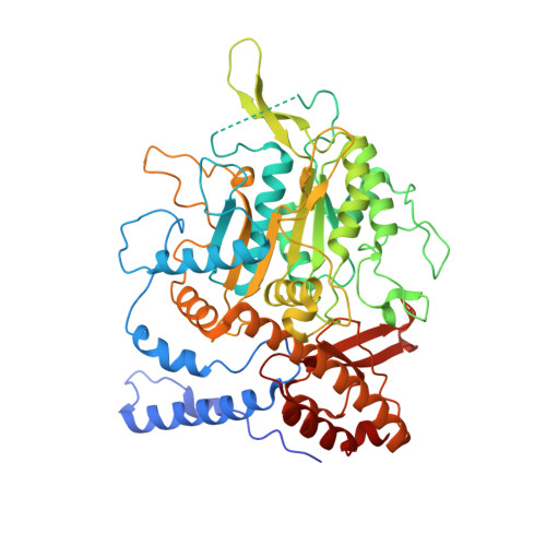
    l -Methionine decarboxylase (MetDC) from Streptomyces sp. 590 is a vitamin B 6 -dependent enzyme and catalyzes the non-oxidative decarboxylation of l -methionine to produce 3-methylthiopropylamine and carbon dioxide. We present here the crystal structures of the ligand-free form of MetDC and of several enzymatic reaction intermediates. Group II amino acid decarboxylases have many residues in common around the active site but the residues surrounding the side chain of the substrate differ. Based on information obtained from the crystal structure, and mutational and biochemical experiments, we propose a key role for Gln64 in determining the substrate specificity of MetDC, and for Tyr421 as the acid catalyst that participates in protonation after the decarboxylation reaction.


  • Organizational Affiliation

    Department of Biofunctional Chemistry, Okayama University, Okayama, Japan.


Macromolecules
Find similar proteins by:  (by identity cutoff)  |  3D Structure
Entity ID: 1
MoleculeChains Sequence LengthOrganismDetailsImage
L-methionine decarboxylase
A, B
557Streptomyces sp. 590 KI-2014Mutation(s): 0 
EC: 4.1.1.57
UniProt
Find proteins for A0A0G4DBU7 (Streptomyces sp. 590 KI-2014)
Explore A0A0G4DBU7 
Go to UniProtKB:  A0A0G4DBU7
Entity Groups  
Sequence Clusters30% Identity50% Identity70% Identity90% Identity95% Identity100% Identity
UniProt GroupA0A0G4DBU7
Sequence Annotations
Expand
  • Reference Sequence
Small Molecules
Ligands 1 Unique
IDChains Name / Formula / InChI Key2D Diagram3D Interactions
G0F (Subject of Investigation/LOI)
Query on G0F

Download Ideal Coordinates CCD File 
C [auth A],
D [auth B]
[6-methyl-4-[(3-methylsulfanylpropylamino)methyl]-5-oxidanyl-pyridin-3-yl]methyl dihydrogen phosphate
C12 H21 N2 O5 P S
AGFAHJKGUOFCGH-UHFFFAOYSA-N
Experimental Data & Validation

Experimental Data

  • Method: X-RAY DIFFRACTION
  • Resolution: 1.80 Å
  • R-Value Free: 0.238 
  • R-Value Work: 0.203 
  • R-Value Observed: 0.204 
  • Space Group: C 1 2 1
Unit Cell:
Length ( Å )Angle ( ˚ )
a = 125.639α = 90
b = 147.813β = 99.47
c = 54.187γ = 90
Software Package:
Software NamePurpose
REFMACrefinement
XDSdata reduction
XDSdata scaling
MOLREPphasing

Structure Validation

View Full Validation Report


Ligand Structure Quality Assessment 


Entry History 

Deposition Data

Revision History  (Full details and data files)

  • Version 1.0: 2021-01-27
    Type: Initial release
  • Version 1.1: 2021-03-03
    Changes: Database references
  • Version 1.2: 2023-11-29
    Changes: Data collection, Database references, Refinement description