7CBB | pdb_00007cbb

Crystal structure of SbnC in the biosynthesis of staphyloferrin B


Experimental Data Snapshot

  • Method: X-RAY DIFFRACTION
  • Resolution: 2.60 Å
  • R-Value Free: 
    0.211 (Depositor), 0.221 (DCC) 
  • R-Value Work: 
    0.197 (Depositor), 0.207 (DCC) 
  • R-Value Observed: 
    0.197 (Depositor) 

wwPDB Validation 3D Report Full Report

Validation slider image for 7CBB

Ligand Structure Quality Assessment 


This is version 1.2 of the entry. See complete history

Literature

Structural and Biochemical Characterization of SbnC as a Representative Type B Siderophore Synthetase.

Tang, J.Ju, Y.Zhou, J.Guo, J.Gu, Q.Xu, J.Zhou, H.

(2020) ACS Chem Biol 15: 2731-2740

  • DOI: https://doi.org/10.1021/acschembio.0c00523
  • Primary Citation Related Structures: 
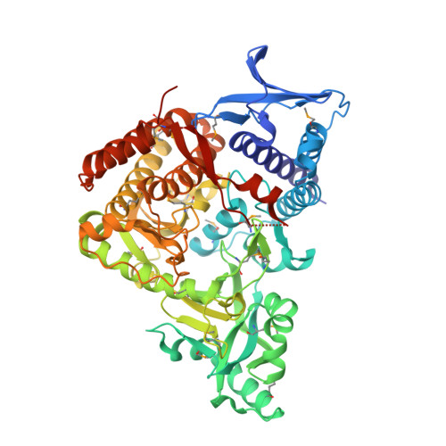
    7CBB

  • PubMed Abstract: 

    Staphyloferrin B is a key siderophore secreted by Staphylococcus aureus to acquire ferric ions from a host during infection, and its biosynthetic pathway has been validated to develop efficient antibacterial agents. Herein, we report the crystal structure of AMP-bound SbnC from S. aureus ( Sa SbnC) as the first representative structure of type B synthetases in the biosynthesis of α-hydroxycarboxylate siderophores. While type B synthetases specifically use α-ketoglutarate (α-KG) as their carboxylic acid substrate, Sa SbnC showed unique structural features in the substrate pocket compared with the type A and C synthetases. Screening of α-KG analogues suggested that the hydrogen-bonding interaction between the α-carbonyl group of α-KG and residue Lys552 is a key determinant for the substrate selectivity of type B synthetases. Interestingly, citrate, the product of the tricarboxylic acid cycle and the substrate of type A synthetases, was found to inhibit the activity of Sa SbnC with an IC 50 value of 83 μM by mimicking α-KG binding, suggesting a potential regulatory role of the tricarboxylic acid cycle, whose activity is under the control of the intracellular iron concentration, to Sa SbnC and other type B synthetases. These results provide critical new information to understand the structure, function, and regulation of type B synthetases in the siderophore-based iron acquisition system employed by a large number of pathogenic microbes.


  • Organizational Affiliation
    • Research Center for Drug Discovery, School of Pharmaceutical Sciences, Sun Yat-sen University, Guangzhou 510006, China.

Macromolecule Content 

  • Total Structure Weight: 137.07 kDa 
  • Atom Count: 9,419 
  • Modeled Residue Count: 1,141 
  • Deposited Residue Count: 1,180 
  • Unique protein chains: 1

Macromolecules

Find similar proteins by:|  3D Structure
Entity ID: 1
MoleculeChains  Sequence LengthOrganismDetailsImage
IucA/IucC family siderophore biosynthesis protein
A, B
590Staphylococcus aureusMutation(s): 0 
Gene Names: sbnCDDL17_05250M1K003_0650
EC: 6.3.2.56
UniProt
Find proteins for Q2G1N1 (Staphylococcus aureus (strain NCTC 8325 / PS 47))
Explore Q2G1N1 
Go to UniProtKB:  Q2G1N1
Entity Groups
Sequence Clusters30% Identity50% Identity70% Identity90% Identity95% Identity100% Identity
UniProt GroupQ2G1N1
Sequence Annotations
Expand
Reference Sequence

Small Molecules

Modified Residues  1 Unique
IDChains TypeFormula2D DiagramParent
MSE
Query on MSE
A, B
L-PEPTIDE LINKINGC5 H11 N O2 SeMET

Experimental Data & Validation

Experimental Data

  • Method: X-RAY DIFFRACTION
  • Resolution: 2.60 Å
  • R-Value Free:  0.211 (Depositor), 0.221 (DCC) 
  • R-Value Work:  0.197 (Depositor), 0.207 (DCC) 
  • R-Value Observed: 0.197 (Depositor) 
Space Group: I 2 2 2
Unit Cell:
Length ( Å )Angle ( ˚ )
a = 140.81α = 90
b = 151.319β = 90
c = 197.08γ = 90
Software Package:
Software NamePurpose
REFMACrefinement
XDSdata reduction
Aimlessdata scaling
AutoSolphasing

Structure Validation

View Full Validation Report


Ligand Structure Quality Assessment 


Entry History 

& Funding Information

Deposition Data


Funding OrganizationLocationGrant Number
National Natural Science Foundation of China (NSFC)China81773636

Revision History  (Full details and data files)

  • Version 1.0: 2020-09-23
    Type: Initial release
  • Version 1.1: 2020-10-28
    Changes: Database references
  • Version 1.2: 2024-10-23
    Changes: Data collection, Database references, Structure summary