7C8E | pdb_00007c8e

Crystal Structure of 14-3-3 epsilon with 9J10 peptide


Experimental Data Snapshot

  • Method: X-RAY DIFFRACTION
  • Resolution: 3.16 Å
  • R-Value Free: 
    0.271 (Depositor), 0.272 (DCC) 
  • R-Value Work: 
    0.222 (Depositor), 0.226 (DCC) 
  • R-Value Observed: 
    0.224 (Depositor) 

Starting Model: experimental
View more details

wwPDB Validation 3D Report Full Report

Validation slider image for 7C8E

Ligand Structure Quality Assessment 


This is version 1.4 of the entry. See complete history

Literature

Target identification for small-molecule discovery in the FOXO3a tumor-suppressor pathway using a biodiverse peptide library.

Emery, A.Hardwick, B.S.Crooks, A.T.Milech, N.Watt, P.M.Mithra, C.Kumar, V.Giridharan, S.Sadasivam, G.Mathivanan, S.Sudhakar, S.Bairy, S.Bharatham, K.Hurakadli, M.A.Prasad, T.K.Kamariah, N.Muellner, M.Coelho, M.Torrance, C.J.McKenzie, G.J.Venkitaraman, A.R.

(2021) Cell Chem Biol 28: 1602-1615.e9

  • DOI: https://doi.org/10.1016/j.chembiol.2021.05.009
  • Primary Citation Related Structures: 
    7C8E

  • PubMed Abstract: 

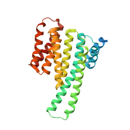
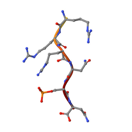
    Genetic screening technologies to identify and validate macromolecular interactions (MMIs) essential for complex pathways remain an important unmet need for systems biology and therapeutics development. Here, we use a library of peptides from diverse prokaryal genomes to screen MMIs promoting the nuclear relocalization of Forkhead Box O3 (FOXO3a), a tumor suppressor more frequently inactivated by post-translational modification than mutation. A hit peptide engages the 14-3-3 family of signal regulators through a phosphorylation-dependent interaction, modulates FOXO3a-mediated transcription, and suppresses cancer cell growth. In a crystal structure, the hit peptide occupies the phosphopeptide-binding groove of 14-3-3ε in a conformation distinct from its natural peptide substrates. A biophysical screen identifies drug-like small molecules that displace the hit peptide from 14-3-3ε, providing starting points for structure-guided development. Our findings exemplify "protein interference," an approach using evolutionarily diverse, natural peptides to rapidly identify, validate, and develop chemical probes against MMIs essential for complex cellular phenotypes.


  • Organizational Affiliation
    • Medical Research Council Cancer Unit, University of Cambridge, Hills Road, Cambridge CB2 0XZ, UK.

Macromolecule Content 

  • Total Structure Weight: 63.51 kDa 
  • Atom Count: 3,735 
  • Modeled Residue Count: 473 
  • Deposited Residue Count: 556 
  • Unique protein chains: 2

Macromolecules

Find similar proteins by:|  3D Structure
Entity ID: 1
MoleculeChains  Sequence LengthOrganismDetailsImage
14-3-3 protein epsilon
A, B
266Homo sapiensMutation(s): 0 
Gene Names: YWHAE
UniProt & NIH Common Fund Data Resources
Find proteins for P62258 (Homo sapiens)
Explore P62258 
Go to UniProtKB:  P62258
PHAROS:  P62258
GTEx:  ENSG00000108953 
Entity Groups
Sequence Clusters30% Identity50% Identity70% Identity90% Identity95% Identity100% Identity
UniProt GroupP62258
Sequence Annotations
Expand
Reference Sequence
Find similar proteins by:  Sequence   |   3D Structure  
Entity ID: 2
MoleculeChains  Sequence LengthOrganismDetailsImage
9J10
C, D
12Streptomyces avermitilis MA-4680 = NBRC 14893Mutation(s): 0 

Small Molecules

Ligands 1 Unique
IDChains Name / Formula / InChI Key2D Diagram3D Interactions
TRS
(Subject of Investigation/LOI)

Query on TRS


Download:Ideal Coordinates CCD File
E [auth B]2-AMINO-2-HYDROXYMETHYL-PROPANE-1,3-DIOL
C4 H12 N O3
LENZDBCJOHFCAS-UHFFFAOYSA-O
Modified Residues  1 Unique
IDChains TypeFormula2D DiagramParent
SEP
Query on SEP
C, D
L-PEPTIDE LINKINGC3 H8 N O6 PSER

Experimental Data & Validation

Experimental Data

  • Method: X-RAY DIFFRACTION
  • Resolution: 3.16 Å
  • R-Value Free:  0.271 (Depositor), 0.272 (DCC) 
  • R-Value Work:  0.222 (Depositor), 0.226 (DCC) 
  • R-Value Observed: 0.224 (Depositor) 
Space Group: P 21 21 21
Unit Cell:
Length ( Å )Angle ( ˚ )
a = 59.47α = 90
b = 83.35β = 90
c = 118.81γ = 90
Software Package:
Software NamePurpose
REFMACrefinement
PDB_EXTRACTdata extraction
MOSFLMdata reduction
SCALAdata scaling
PHASERphasing

Structure Validation

View Full Validation Report


Ligand Structure Quality Assessment 


Entry History 

& Funding Information

Deposition Data


Funding OrganizationLocationGrant Number
Department of Biotechnology (DBT, India)India--

Revision History  (Full details and data files)

  • Version 1.0: 2021-06-02
    Type: Initial release
  • Version 1.1: 2021-06-30
    Changes: Database references
  • Version 1.2: 2021-12-08
    Changes: Database references
  • Version 1.3: 2023-11-29
    Changes: Data collection, Refinement description
  • Version 1.4: 2024-10-09
    Changes: Structure summary