7C7S

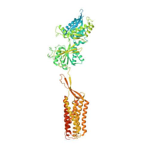
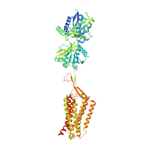
Cryo-EM structure of the CGP54626-bound human GABA(B) receptor in inactive state.


Experimental Data Snapshot

  • Method: ELECTRON MICROSCOPY
  • Resolution: 2.90 Å
  • Aggregation State: PARTICLE 
  • Reconstruction Method: SINGLE PARTICLE 

wwPDB Validation   3D Report Full Report


This is version 1.2 of the entry. See complete history


Literature

Cryo-EM structures of inactive and active GABABreceptor.

Mao, C.Shen, C.Li, C.Shen, D.D.Xu, C.Zhang, S.Zhou, R.Shen, Q.Chen, L.N.Jiang, Z.Liu, J.Zhang, Y.

(2020) Cell Res 30: 564-573

  • DOI: https://doi.org/10.1038/s41422-020-0350-5
  • Primary Citation of Related Structures:  
    7C7Q, 7C7S

  • PubMed Abstract: 

    Metabotropic GABA B G protein-coupled receptor functions as a mandatory heterodimer of GB1 and GB2 subunits and mediates inhibitory neurotransmission in the central nervous system. Each subunit is composed of the extracellular Venus flytrap (VFT) domain and transmembrane (TM) domain. Here we present cryo-EM structures of full-length human heterodimeric GABA B receptor in the antagonist-bound inactive state and in the active state complexed with an agonist and a positive allosteric modulator in the presence of G i1 protein at a resolution range of 2.8-3.0 Å. Our structures reveal that agonist binding stabilizes the closure of GB1 VFT, which in turn triggers a rearrangement of TM interfaces between the two subunits from TM3-TM5/TM3-TM5 in the inactive state to TM6/TM6 in the active state and finally induces the opening of intracellular loop 3 and synergistic shifting of TM3, 4 and 5 helices in GB2 TM domain to accommodate the α5-helix of G i1 . We also observed that the positive allosteric modulator anchors at the dimeric interface of TM domains. These results provide a structural framework for understanding class C GPCR activation and a rational template for allosteric modulator design targeting the dimeric interface of GABA B receptor.


  • Organizational Affiliation

    Department of Biophysics, and Department of Pathology of Sir Run Run Shaw Hospital, Zhejiang University School of Medicine, Hangzhou, 310058, Zhejiang, China.


Macromolecules
Find similar proteins by:  (by identity cutoff)  |  3D Structure
Entity ID: 1
MoleculeChains Sequence LengthOrganismDetailsImage
Gamma-aminobutyric acid type B receptor subunit 1937Homo sapiensMutation(s): 0 
Gene Names: GABBR1GPRC3A
Membrane Entity: Yes 
UniProt & NIH Common Fund Data Resources
Find proteins for Q9UBS5 (Homo sapiens)
Explore Q9UBS5 
Go to UniProtKB:  Q9UBS5
PHAROS:  Q9UBS5
GTEx:  ENSG00000204681 
Entity Groups  
Sequence Clusters30% Identity50% Identity70% Identity90% Identity95% Identity100% Identity
UniProt GroupQ9UBS5
Glycosylation
Glycosylation Sites: 5Go to GlyGen: Q9UBS5-1
Sequence Annotations
Expand
  • Reference Sequence
Find similar proteins by:  (by identity cutoff)  |  3D Structure
Entity ID: 2
MoleculeChains Sequence LengthOrganismDetailsImage
Gamma-aminobutyric acid type B receptor subunit 2808Homo sapiensMutation(s): 0 
Gene Names: GABBR2GPR51GPRC3B
Membrane Entity: Yes 
UniProt & NIH Common Fund Data Resources
Find proteins for O75899 (Homo sapiens)
Explore O75899 
Go to UniProtKB:  O75899
PHAROS:  O75899
GTEx:  ENSG00000136928 
Entity Groups  
Sequence Clusters30% Identity50% Identity70% Identity90% Identity95% Identity100% Identity
UniProt GroupO75899
Glycosylation
Glycosylation Sites: 3Go to GlyGen: O75899-1
Sequence Annotations
Expand
  • Reference Sequence
Small Molecules
Ligands 2 Unique
IDChains Name / Formula / InChI Key2D Diagram3D Interactions
2BV (Subject of Investigation/LOI)
Query on 2BV

Download Ideal Coordinates CCD File 
H [auth A](R)-(cyclohexylmethyl)[(2S)-3-{[(1S)-1-(3,4-dichlorophenyl)ethyl]amino}-2-hydroxypropyl]phosphinic acid
C18 H28 Cl2 N O3 P
JGGVBBYJRQOPPA-BBRMVZONSA-N
NAG
Query on NAG

Download Ideal Coordinates CCD File 
C [auth A]
D [auth A]
E [auth A]
F [auth A]
G [auth A]
C [auth A],
D [auth A],
E [auth A],
F [auth A],
G [auth A],
I [auth B],
J [auth B],
K [auth B]
2-acetamido-2-deoxy-beta-D-glucopyranose
C8 H15 N O6
OVRNDRQMDRJTHS-FMDGEEDCSA-N
Experimental Data & Validation

Experimental Data

  • Method: ELECTRON MICROSCOPY
  • Resolution: 2.90 Å
  • Aggregation State: PARTICLE 
  • Reconstruction Method: SINGLE PARTICLE 
EM Software:
TaskSoftware PackageVersion
RECONSTRUCTIONRELION3.0-beta2
MODEL REFINEMENTPHENIX1.16

Structure Validation

View Full Validation Report


Entry History & Funding Information

Deposition Data


Funding OrganizationLocationGrant Number
National Natural Science Foundation of China (NSFC)China81922071
Ministry of Science and Technology (MoST, China)China2018YFA0507003

Revision History  (Full details and data files)

  • Version 1.0: 2020-07-01
    Type: Initial release
  • Version 1.1: 2020-07-22
    Changes: Database references
  • Version 1.2: 2020-07-29
    Type: Remediation
    Reason: Carbohydrate remediation
    Changes: Data collection, Derived calculations, Structure summary