7C4C

The crystal structure of Trypanosoma brucei RNase D : GMP complex


Experimental Data Snapshot

  • Method: X-RAY DIFFRACTION
  • Resolution: 2.27 Å
  • R-Value Free: 0.239 
  • R-Value Work: 0.192 
  • R-Value Observed: 0.195 

wwPDB Validation   3D Report Full Report


Ligand Structure Quality Assessment 


This is version 1.0 of the entry. See complete history


Literature

Structural basis for guide RNA trimming by RNase D ribonuclease in Trypanosoma brucei.

Gao, Y.Liu, H.Zhang, C.Su, S.Chen, Y.Chen, X.Li, Y.Shao, Z.Zhang, Y.Shao, Q.Li, J.Huang, Z.Ma, J.Gan, J.

(2021) Nucleic Acids Res 49: 568-583

  • DOI: https://doi.org/10.1093/nar/gkaa1197
  • Primary Citation of Related Structures:  
    7C42, 7C43, 7C45, 7C47, 7C4B, 7C4C

  • PubMed Abstract: 

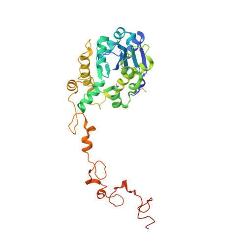
    Infection with kinetoplastid parasites, including Trypanosoma brucei (T. brucei), Trypanosoma cruzi (T. cruzi) and Leishmania can cause serious disease in humans. Like other kinetoplastid species, mRNAs of these disease-causing parasites must undergo posttranscriptional editing in order to be functional. mRNA editing is directed by gRNAs, a large group of small RNAs. Similar to mRNAs, gRNAs are also precisely regulated. In T. brucei, overexpression of RNase D ribonuclease (TbRND) leads to substantial reduction in the total gRNA population and subsequent inhibition of mRNA editing. However, the mechanisms regulating gRNA binding and cleavage by TbRND are not well defined. Here, we report a thorough structural study of TbRND. Besides Apo- and NMP-bound structures, we also solved one TbRND structure in complexed with single-stranded RNA. In combination with mutagenesis and in vitro cleavage assays, our structures indicated that TbRND follows the conserved two-cation-assisted mechanism in catalysis. TbRND is a unique RND member, as it contains a ZFD domain at its C-terminus. In addition to T. brucei, our studies also advanced our understanding on the potential gRNA degradation pathway in T. cruzi, Leishmania, as well for as other disease-associated parasites expressing ZFD-containing RNDs.


  • Organizational Affiliation

    Shanghai Public Health Clinical Center, State Key Laboratory of Genetic Engineering, Collaborative Innovation Center of Genetics and Development, Department of Physiology and Biophysics, School of Life Sciences, Fudan University, Shanghai 200438, China.


Macromolecules
Find similar proteins by:  (by identity cutoff)  |  3D Structure
Entity ID: 1
MoleculeChains Sequence LengthOrganismDetailsImage
CCHC-type domain-containing protein351Trypanosoma brucei brucei TREU927Mutation(s): 0 
Gene Names: Tb09.211.3670
UniProt
Find proteins for Q38DE2 (Trypanosoma brucei brucei (strain 927/4 GUTat10.1))
Explore Q38DE2 
Go to UniProtKB:  Q38DE2
Entity Groups  
Sequence Clusters30% Identity50% Identity70% Identity90% Identity95% Identity100% Identity
UniProt GroupQ38DE2
Sequence Annotations
Expand
  • Reference Sequence
Small Molecules
Ligands 3 Unique
IDChains Name / Formula / InChI Key2D Diagram3D Interactions
5GP (Subject of Investigation/LOI)
Query on 5GP

Download Ideal Coordinates CCD File 
N [auth A],
O [auth A],
P [auth A],
Q [auth A]
GUANOSINE-5'-MONOPHOSPHATE
C10 H14 N5 O8 P
RQFCJASXJCIDSX-UUOKFMHZSA-N
ZN (Subject of Investigation/LOI)
Query on ZN

Download Ideal Coordinates CCD File 
B [auth A]
C [auth A]
D [auth A]
E [auth A]
F [auth A]
B [auth A],
C [auth A],
D [auth A],
E [auth A],
F [auth A],
G [auth A]
ZINC ION
Zn
PTFCDOFLOPIGGS-UHFFFAOYSA-N
MN (Subject of Investigation/LOI)
Query on MN

Download Ideal Coordinates CCD File 
H [auth A]
I [auth A]
J [auth A]
K [auth A]
L [auth A]
H [auth A],
I [auth A],
J [auth A],
K [auth A],
L [auth A],
M [auth A]
MANGANESE (II) ION
Mn
WAEMQWOKJMHJLA-UHFFFAOYSA-N
Modified Residues  1 Unique
IDChains TypeFormula2D DiagramParent
MSE
Query on MSE
A
L-PEPTIDE LINKINGC5 H11 N O2 SeMET
Experimental Data & Validation

Experimental Data

  • Method: X-RAY DIFFRACTION
  • Resolution: 2.27 Å
  • R-Value Free: 0.239 
  • R-Value Work: 0.192 
  • R-Value Observed: 0.195 
  • Space Group: P 21 21 21
Unit Cell:
Length ( Å )Angle ( ˚ )
a = 57.169α = 90
b = 75.001β = 90
c = 101.409γ = 90
Software Package:
Software NamePurpose
PHENIXrefinement
HKL-2000data scaling
PDB_EXTRACTdata extraction
HKL-3000data reduction
HKL2Mapphasing

Structure Validation

View Full Validation Report


Ligand Structure Quality Assessment 


Entry History & Funding Information

Deposition Data


Funding OrganizationLocationGrant Number
Ministry of Science and Technology (MoST, China)China2019FY101500

Revision History  (Full details and data files)

  • Version 1.0: 2021-04-07
    Type: Initial release