7C1M

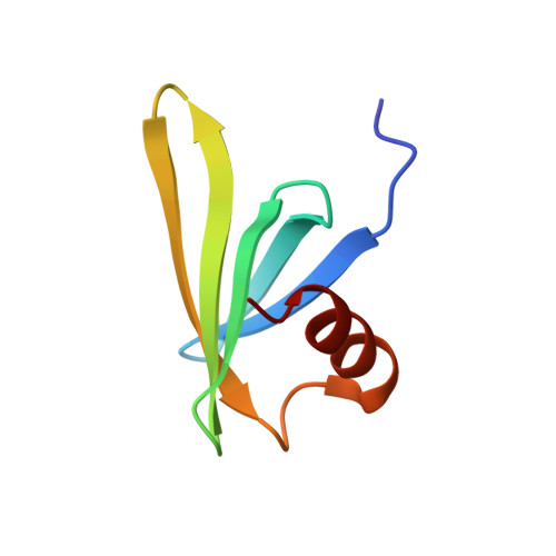
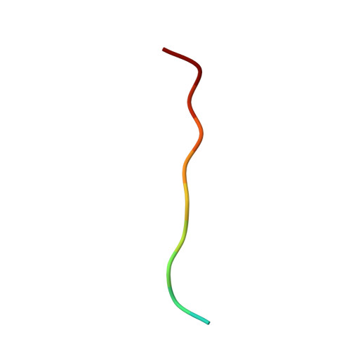
Complex structure of tyrosinated alpha-tubulin carboxy-terminal peptide and A1aY1 binder


Experimental Data Snapshot

  • Method: SOLUTION NMR
  • Conformers Calculated: 200 
  • Conformers Submitted: 20 
  • Selection Criteria: structures with the lowest energy 

wwPDB Validation   3D Report Full Report


This is version 1.3 of the entry. See complete history


Literature

Genetically encoded live-cell sensor for tyrosinated microtubules.

Kesarwani, S.Lama, P.Chandra, A.Reddy, P.P.Jijumon, A.S.Bodakuntla, S.Rao, B.M.Janke, C.Das, R.Sirajuddin, M.

(2020) J Cell Biol 219

  • DOI: https://doi.org/10.1083/jcb.201912107
  • Primary Citation of Related Structures:  
    7C1M

  • PubMed Abstract: 

    Microtubule cytoskeleton exists in various biochemical forms in different cells due to tubulin posttranslational modifications (PTMs). Tubulin PTMs are known to affect microtubule stability, dynamics, and interaction with MAPs and motors in a specific manner, widely known as tubulin code hypothesis. At present, there exists no tool that can specifically mark tubulin PTMs in living cells, thus severely limiting our understanding of their dynamics and cellular functions. Using a yeast display library, we identified a binder against terminal tyrosine of α-tubulin, a unique PTM site. Extensive characterization validates the robustness and nonperturbing nature of our binder as tyrosination sensor, a live-cell tubulin nanobody specific towards tyrosinated microtubules. Using this sensor, we followed nocodazole-, colchicine-, and vincristine-induced depolymerization events of tyrosinated microtubules in real time and found each distinctly perturbs the microtubule polymer. Together, our work describes a novel tyrosination sensor and its potential applications to study the dynamics of microtubule and their PTM processes in living cells.


  • Organizational Affiliation

    Centre for Cardiovascular Biology and Diseases, Institute for Stem Cell Science and Regenerative Medicine, Gandhi Krishi Vigyan Kendra Campus, Bangalore, India.


Macromolecules
Find similar proteins by:  (by identity cutoff)  |  3D Structure
Entity ID: 1
MoleculeChains Sequence LengthOrganismDetailsImage
Nanobody binder from SSO7d library67Saccharolobus solfataricus 98/2Mutation(s): 0 
Entity Groups  
Sequence Clusters30% Identity50% Identity70% Identity90% Identity95% Identity100% Identity
Sequence Annotations
Expand
  • Reference Sequence

Find similar proteins by:  Sequence   |   3D Structure  

Entity ID: 2
MoleculeChains Sequence LengthOrganismDetailsImage
Carboxy-terminal peptide from tyrosinated alpha-tubulin12Homo sapiensMutation(s): 0 
EC: 3.6.5
UniProt & NIH Common Fund Data Resources
Find proteins for Q71U36 (Homo sapiens)
Explore Q71U36 
Go to UniProtKB:  Q71U36
PHAROS:  Q71U36
GTEx:  ENSG00000167552 
Entity Groups  
Sequence Clusters30% Identity50% Identity70% Identity90% Identity95% Identity100% Identity
UniProt GroupQ71U36
Sequence Annotations
Expand
  • Reference Sequence
Experimental Data & Validation

Experimental Data

  • Method: SOLUTION NMR
  • Conformers Calculated: 200 
  • Conformers Submitted: 20 
  • Selection Criteria: structures with the lowest energy 

Structure Validation

View Full Validation Report


Entry History & Funding Information

Deposition Data


Funding OrganizationLocationGrant Number
Department of Biotechnology (DBT, India)IndiaDBT/PR5081/INF/156/2012

Revision History  (Full details and data files)

  • Version 1.0: 2020-09-02
    Type: Initial release
  • Version 1.1: 2020-10-14
    Changes: Database references
  • Version 1.2: 2023-06-14
    Changes: Database references, Other
  • Version 1.3: 2024-05-15
    Changes: Data collection, Database references