7BZ0 | pdb_00007bz0

complex structure of alginate lyase AlyF-OU02 with G6


Experimental Data Snapshot

  • Method: X-RAY DIFFRACTION
  • Resolution: 1.80 Å
  • R-Value Free: 
    0.236 (Depositor), 0.238 (DCC) 
  • R-Value Work: 
    0.199 (Depositor), 0.199 (DCC) 
  • R-Value Observed: 
    0.201 (Depositor) 

Starting Model: experimental
View more details

wwPDB Validation 3D Report Full Report

Validation slider image for 7BZ0

This is version 1.1 of the entry. See complete history

Literature

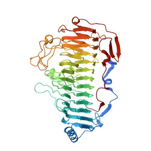
Structural insights into the substrate-binding cleft of AlyF reveal the first long-chain alginate-binding mode.

Zhang, K.Liu, T.Liu, W.Lyu, Q.

(2021) Acta Crystallogr D Struct Biol 77: 336-346

  • DOI: https://doi.org/10.1107/S205979832100005X
  • Primary Citation Related Structures: 
    7BZ0

  • PubMed Abstract: 

    The products of alginate degradation, alginate oligosaccharides (AOS), have potential applications in many areas, including functional foods and marine drugs. Enzyme-based approaches using alginate lyases have advantages in the preparation of well defined AOS and have attracted much attention in recent years. However, a lack of structural insight into the whole substrate-binding cleft for most known alginate lyases severely hampers their application in the industrial generation of well defined AOS. To solve this issue, AlyF was co-crystallized with the long alginate oligosaccharide G6 (L-hexaguluronic acid hexasodium salt), which is the longest bound substrate in all solved alginate lyase complex structures. AlyF formed interactions with G6 from subsites -3 to +3 without additional substrate-binding site interactions, suggesting that the substrate-binding cleft of AlyF was fully occupied by six sugars, which was further confirmed by isothermal titration calorimetry and differential scanning calorimetry analyses. More importantly, a combination of structural comparisons and mutagenetic analyses determined that three key loops (loop 1, Lys215-Glu236; loop 2, Gln402-Ile416; loop 3, Arg334-Gly348) mainly function in binding long substrates (degree of polymerization of >4). The potential flexibility of loop 1 and loop 2 might enable the substrate to continue to enter the cleft after binding to subsites +1 to +3; loop 3 stabilizes and orients the substrate at subsites -2 and -3. Taken together, these results provide the first possible alginate lyase-substrate binding profile for long-chain alginates, facilitating the rational design of new enzymes for industrial purposes.


  • Organizational Affiliation
    • MOE Key Laboratory of Marine Genetics and Breeding, College of Marine Life Sciences, Ocean University of China, Qingdao 266003, People's Republic of China.

Macromolecule Content 

  • Total Structure Weight: 60.14 kDa 
  • Atom Count: 4,131 
  • Modeled Residue Count: 496 
  • Deposited Residue Count: 536 
  • Unique protein chains: 1

Macromolecules

Find similar proteins by:|  3D Structure
Entity ID: 1
MoleculeChains  Sequence LengthOrganismDetailsImage
Alginate lyase AlyF-OU02536Vibrio splendidusMutation(s): 0 
EC: 4.2.2

Oligosaccharides

Help  
Entity ID: 2
MoleculeChains Length2D Diagram GlycosylationD Interactions
alpha-L-gulopyranuronic acid-(1-4)-alpha-L-gulopyranuronic acid-(1-4)-alpha-L-gulopyranuronic acid-(1-4)-alpha-L-gulopyranuronic acid-(1-4)-alpha-L-gulopyranuronic acid-(1-4)-alpha-L-gulopyranuronic acid
B
6N/A
Glycosylation Resources
GlyTouCan: G84972FO
GlyCosmos: G84972FO

Small Molecules

Ligands 1 Unique
IDChains Name / Formula / InChI Key2D Diagram3D Interactions
CA

Query on CA


Download:Ideal Coordinates CCD File
C [auth A]CALCIUM ION
Ca
BHPQYMZQTOCNFJ-UHFFFAOYSA-N

Experimental Data & Validation

Experimental Data

  • Method: X-RAY DIFFRACTION
  • Resolution: 1.80 Å
  • R-Value Free:  0.236 (Depositor), 0.238 (DCC) 
  • R-Value Work:  0.199 (Depositor), 0.199 (DCC) 
  • R-Value Observed: 0.201 (Depositor) 
Space Group: P 61
Unit Cell:
Length ( Å )Angle ( ˚ )
a = 105.983α = 90
b = 105.983β = 90
c = 101.665γ = 120
Software Package:
Software NamePurpose
REFMACrefinement
PDB_EXTRACTdata extraction
HKL-2000data reduction
HKL-2000data scaling
PHASERphasing

Structure Validation

View Full Validation Report


Entry History 

& Funding Information

Deposition Data


Funding OrganizationLocationGrant Number
National Natural Science Foundation of China (NSFC)China41706151

Revision History  (Full details and data files)

  • Version 1.0: 2021-03-10
    Type: Initial release
  • Version 1.1: 2023-11-29
    Changes: Data collection, Database references, Refinement description