7BVB

Crystal structure of UDP-N-acetylmuramic Acid L-alanine ligase (MurC) from Mycobacterium bovis in complex with UDP-N-acetylglucosamine


Experimental Data Snapshot

  • Method: X-RAY DIFFRACTION
  • Resolution: 3.19 Å
  • R-Value Free: 0.247 
  • R-Value Work: 0.195 
  • R-Value Observed: 0.198 

wwPDB Validation   3D Report Full Report


Ligand Structure Quality Assessment 


This is version 1.3 of the entry. See complete history


Literature

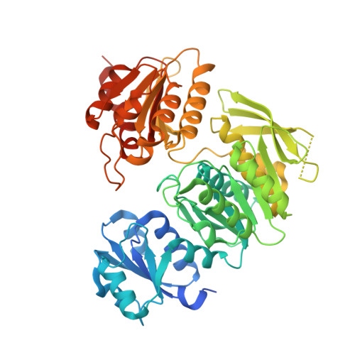
Crystal structures of UDP-N-acetylmuramic acid L-alanine ligase (MurC) from Mycobacterium bovis with and without UDP-N-acetylglucosamine.

Seo, P.W.Park, S.Y.Hofmann, A.Kim, J.S.

(2021) Acta Crystallogr D Struct Biol 77: 618-627

  • DOI: https://doi.org/10.1107/S2059798321002199
  • Primary Citation of Related Structures:  
    7BVA, 7BVB

  • PubMed Abstract: 

    Peptidoglycan comprises repeating units of N-acetylmuramic acid, N-acetylglucosamine and short cross-linking peptides. After the conversion of UDP-N-acetylglucosamine (UNAG) to UDP-N-acetylmuramic acid (UNAM) by the MurA and MurB enzymes, an amino acid is added to UNAM by UDP-N-acetylmuramic acid L-alanine ligase (MurC). As peptidoglycan is an essential component of the bacterial cell wall, the enzymes involved in its biosynthesis represent promising targets for the development of novel antibacterial drugs. Here, the crystal structure of Mycobacterium bovis MurC (MbMurC) is reported, which exhibits a three-domain architecture for the binding of UNAM, ATP and an amino acid as substrates, with a nickel ion at the domain interface. The ATP-binding loop adopts a conformation that is not seen in other MurCs. In the UNAG-bound structure of MbMurC, the substrate mimic interacts with the UDP-binding domain of MbMurC, which does not invoke rearrangement of the three domains. Interestingly, the glycine-rich loop of the UDP-binding domain of MbMurC interacts through hydrogen bonds with the glucose moiety of the ligand, but not with the pyrophosphate moiety. These findings suggest that UNAG analogs might serve as potential candidates for neutralizing the catalytic activity of bacterial MurC.


  • Organizational Affiliation

    Department of Chemistry, Chonnam National University, Gwangju 61186, Republic of Korea.


Macromolecules
Find similar proteins by:  (by identity cutoff)  |  3D Structure
Entity ID: 1
MoleculeChains Sequence LengthOrganismDetailsImage
UDP-N-acetylmuramate--L-alanine ligase
A, B
519Mycobacterium tuberculosis variant bovis AF2122/97Mutation(s): 0 
Gene Names: murCBQ2027_MB2176C
EC: 6.3.2.8
UniProt
Find proteins for P65473 (Mycobacterium bovis (strain ATCC BAA-935 / AF2122/97))
Explore P65473 
Go to UniProtKB:  P65473
Entity Groups  
Sequence Clusters30% Identity50% Identity70% Identity90% Identity95% Identity100% Identity
UniProt GroupP65473
Sequence Annotations
Expand
  • Reference Sequence
Experimental Data & Validation

Experimental Data

  • Method: X-RAY DIFFRACTION
  • Resolution: 3.19 Å
  • R-Value Free: 0.247 
  • R-Value Work: 0.195 
  • R-Value Observed: 0.198 
  • Space Group: P 1 21 1
Unit Cell:
Length ( Å )Angle ( ˚ )
a = 65.285α = 90
b = 75.762β = 105.92
c = 104.679γ = 90
Software Package:
Software NamePurpose
PHENIXrefinement
HKL-2000data scaling
PDB_EXTRACTdata extraction
HKL-2000data reduction
PHENIXphasing

Structure Validation

View Full Validation Report


Ligand Structure Quality Assessment 


Entry History & Funding Information

Deposition Data


Funding OrganizationLocationGrant Number
National Research Foundation (NRF, Korea)Korea, Republic OfNRF-2019R1I1A3A01061866

Revision History  (Full details and data files)

  • Version 1.0: 2020-04-22
    Type: Initial release
  • Version 1.1: 2021-05-05
    Changes: Database references
  • Version 1.2: 2021-05-19
    Changes: Database references
  • Version 1.3: 2023-11-29
    Changes: Data collection, Database references, Refinement description