7BUY

The crystal structure of COVID-19 main protease in complex with carmofur


Experimental Data Snapshot

  • Method: X-RAY DIFFRACTION
  • Resolution: 1.60 Å
  • R-Value Free: 0.201 
  • R-Value Work: 0.179 
  • R-Value Observed: 0.180 

wwPDB Validation   3D Report Full Report


Ligand Structure Quality Assessment 


This is version 1.3 of the entry. See complete history


Literature

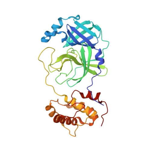
Structural basis for the inhibition of SARS-CoV-2 main protease by antineoplastic drug carmofur.

Jin, Z.Zhao, Y.Sun, Y.Zhang, B.Wang, H.Wu, Y.Zhu, Y.Zhu, C.Hu, T.Du, X.Duan, Y.Yu, J.Yang, X.Yang, X.Yang, K.Liu, X.Guddat, L.W.Xiao, G.Zhang, L.Yang, H.Rao, Z.

(2020) Nat Struct Mol Biol 27: 529-532

  • DOI: https://doi.org/10.1038/s41594-020-0440-6
  • Primary Citation of Related Structures:  
    7BUY

  • PubMed Abstract: 
  • The antineoplastic drug carmofur is shown to inhibit the SARS-CoV-2 main protease (M pro ). Here, the X-ray crystal structure of M pro in complex with carmofur reveals that the carbonyl reactive group of carmofur is covalently bound to catalytic Cys145, whereas its fatty acid tail occupies the hydrophobic S2 subsite ...

    The antineoplastic drug carmofur is shown to inhibit the SARS-CoV-2 main protease (M pro ). Here, the X-ray crystal structure of M pro in complex with carmofur reveals that the carbonyl reactive group of carmofur is covalently bound to catalytic Cys145, whereas its fatty acid tail occupies the hydrophobic S2 subsite. Carmofur inhibits viral replication in cells (EC 50  = 24.30 μM) and is a promising lead compound to develop new antiviral treatment for COVID-19.


    Organizational Affiliation

    State Key Laboratory of Medicinal Chemical Biology, Frontiers Science Center for Cell Response, College of Life Sciences, College of Pharmacy, Nankai University, Tianjin, China.


Macromolecules
Find similar proteins by:  (by identity cutoff)  |  3D Structure
Entity ID: 1
MoleculeChainsSequence LengthOrganismDetailsImage
3C-like proteinaseA306Severe acute respiratory syndrome coronavirus 2Mutation(s): 0 
Gene Names: rep1a-1b
EC: 3.4.22.69 (PDB Primary Data), 3.4.19.12 (UniProt), 3.4.22 (UniProt), 2.7.7.48 (UniProt), 3.6.4.12 (UniProt), 3.6.4.13 (UniProt), 3.1.13 (UniProt), 4.6.1 (UniProt), 2.1.1.57 (UniProt)
UniProt
Find proteins for P0DTD1 (Severe acute respiratory syndrome coronavirus 2)
Explore P0DTD1 
Go to UniProtKB:  P0DTD1
Entity Groups  
Sequence Clusters30% Identity50% Identity70% Identity90% Identity95% Identity100% Identity
UniProt GroupP0DTD1
Protein Feature View
Expand
  • Reference Sequence
Experimental Data & Validation

Experimental Data

  • Method: X-RAY DIFFRACTION
  • Resolution: 1.60 Å
  • R-Value Free: 0.201 
  • R-Value Work: 0.179 
  • R-Value Observed: 0.180 
  • Space Group: C 1 2 1
Unit Cell:
Length ( Å )Angle ( ˚ )
a = 98.028α = 90
b = 81.652β = 114.88
c = 51.639γ = 90
Software Package:
Software NamePurpose
PHENIXrefinement
XSCALEdata scaling
PDB_EXTRACTdata extraction
XDSdata reduction
PHASERphasing

Structure Validation

View Full Validation Report


Ligand Structure Quality Assessment 


Entry History & Funding Information

Deposition Data


Funding OrganizationLocationGrant Number
National Natural Science Foundation of China (NSFC)China81520108019
National Natural Science Foundation of China (NSFC)China813300237

Revision History  (Full details and data files)

  • Version 1.0: 2020-04-29
    Type: Initial release
  • Version 1.1: 2020-05-20
    Changes: Database references
  • Version 1.2: 2020-06-24
    Changes: Database references
  • Version 1.3: 2021-02-17
    Changes: Database references, Source and taxonomy, Structure summary