Discovery of novel histone lysine methyltransferase G9a/GLP (EHMT2/1) inhibitors: Design, synthesis, and structure-activity relationships of 2,4-diamino-6-methylpyrimidines.
Katayama, K., Ishii, K., Tsuda, E., Yotsumoto, K., Hiramoto, K., Suzuki, M., Yasumatsu, I., Igarashi, W., Torihata, M., Ishiyama, T., Katagiri, T.(2020) Bioorg Med Chem Lett 30: 127475-127475
- PubMed: 32781218 Search on PubMed
- DOI: https://doi.org/10.1016/j.bmcl.2020.127475
- Primary Citation Related Structures:
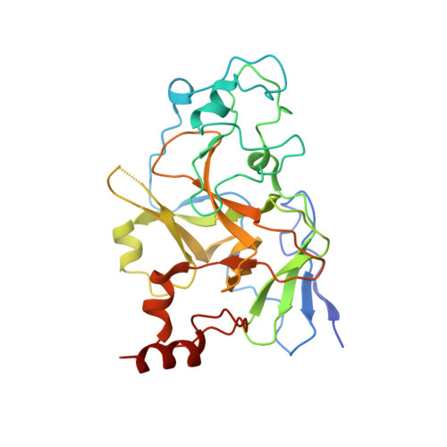
7BTV, 7BUC - PubMed Abstract:
The discovery and optimization of a novel series of G9a/GLP (EHMT2/1) inhibitors are described. Starting from known G9a/GLP inhibitor 5, efforts to explore the structure-activity relationship and optimize drug properties led to a novel compound 13, the side chain of which was converted to tetrahydroazepine. Compound 13 showed increased G9a/GLP inhibitory activity compared with compound 5. In addition, compound 13 exhibited improved human ether-a-go-go related gene (hERG) inhibitory activity over compound 5 and also improved pharmacokinetic profile in mice (oral bioavailability: 17 to 40%). Finally, the co-crystal structure of G9a in complex with compound 13 provides the basis for the further development of tetrahydroazepine-based G9a/GLP inhibitors.
- R&D Division, Daiichi Sankyo Co., Ltd., 1-2-58 Hiromachi, Shinagawa-ku, Tokyo 140-8710, Japan. Electronic address: katayama.katsushi.ne@daiichisankyo.co.jp.
Organizational Affiliation: