7BQF | pdb_00007bqf

Dimerization of SAV1 WW tandem


Experimental Data Snapshot

  • Method: X-RAY DIFFRACTION
  • Resolution: 1.70 Å
  • R-Value Free: 
    0.220 (Depositor), 0.220 (DCC) 
  • R-Value Work: 
    0.196 (Depositor), 0.200 (DCC) 
  • R-Value Observed: 
    0.198 (Depositor) 

Starting Models: experimental
View more details

wwPDB Validation   3D Report Full Report


Ligand Structure Quality Assessment 


This is version 1.2 of the entry. See complete history


Literature

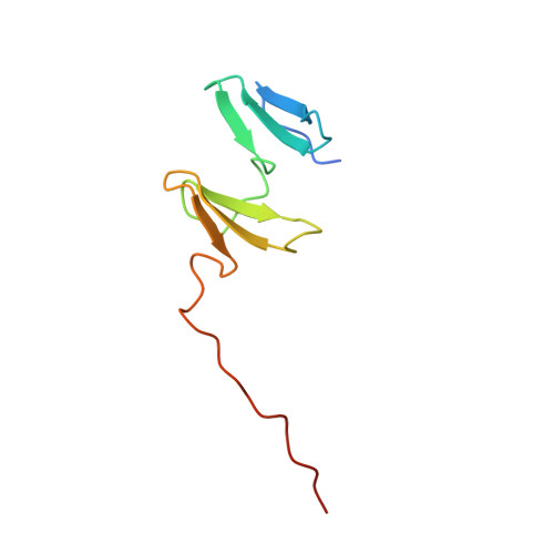
A WW Tandem-Mediated Dimerization Mode of SAV1 Essential for Hippo Signaling.

Lin, Z.Xie, R.Guan, K.Zhang, M.

(2020) Cell Rep 32: 108118-108118

  • DOI: https://doi.org/10.1016/j.celrep.2020.108118
  • Primary Citation of Related Structures:  
    7BQF, 7BQG

  • PubMed Abstract: 

    The canonical mammalian Hippo pathway contains a core kinase signaling cascade requiring upstream MST to form a stable complex with SAV1 in order to phosphorylate the downstream LATS/MOB complex. Though SAV1 dimerization is essential for the trans-activation of MST, the molecular mechanism underlying SAV1 dimerization is unclear. Here, we discover that the SAV1 WW tandem containing a short Pro-rich extension immediately following the WW tandem (termed as "WW12ex") forms a highly stable homodimer. The crystal structure of SAV1 WW12ex reveals that the Pro-rich extension of one subunit binds to both WW domains from the other subunit. Thus, SAV1 WW12ex forms a domain-swapped dimer instead of a WW2 homodimerization-mediated dimer. The WW12ex-mediated dimerization of SAV1 is required for the MST/SAV1 complex assembly and MST kinase activation. Finally, we show that several cancer-related SAV1 variants disrupt SAV1 dimer formation, and thus, these mutations may impair the tumor-suppression activity of SAV1.


  • Organizational Affiliation
    • Division of Life Science, State Key Laboratory of Molecular Neuroscience, Hong Kong University of Science and Technology, Clear Water Bay, Kowloon, Hong Kong, China.

Macromolecules
Find similar proteins by:  (by identity cutoff)  |  3D Structure
Entity ID: 1
MoleculeChains Sequence LengthOrganismDetailsImage
Protein salvador homolog 192Mus musculusMutation(s): 0 
Gene Names: Sav1Ww45Wwp3
UniProt
Find proteins for Q8VEB2 (Mus musculus)
Explore Q8VEB2 
Go to UniProtKB:  Q8VEB2
Entity Groups  
Sequence Clusters30% Identity50% Identity70% Identity90% Identity95% Identity100% Identity
UniProt GroupQ8VEB2
Sequence Annotations
Expand
  • Reference Sequence
Small Molecules
Ligands 1 Unique
IDChains Name / Formula / InChI Key2D Diagram3D Interactions
DIO (Subject of Investigation/LOI)
Query on DIO

Download Ideal Coordinates CCD File 
B [auth A]1,4-DIETHYLENE DIOXIDE
C4 H8 O2
RYHBNJHYFVUHQT-UHFFFAOYSA-N
Experimental Data & Validation

Experimental Data

  • Method: X-RAY DIFFRACTION
  • Resolution: 1.70 Å
  • R-Value Free:  0.220 (Depositor), 0.220 (DCC) 
  • R-Value Work:  0.196 (Depositor), 0.200 (DCC) 
  • R-Value Observed: 0.198 (Depositor) 
Space Group: P 31 2 1
Unit Cell:
Length ( Å )Angle ( ˚ )
a = 46.715α = 90
b = 46.715β = 90
c = 83.151γ = 120
Software Package:
Software NamePurpose
PHENIXrefinement
HKL-3000data scaling
PHASERphasing
HKL-3000data reduction

Structure Validation

View Full Validation Report


Ligand Structure Quality Assessment 


Entry History & Funding Information

Deposition Data


Funding OrganizationLocationGrant Number
The University Grants Committee, Research Grants Council (RGC)Hong Kong--

Revision History  (Full details and data files)

  • Version 1.0: 2020-09-23
    Type: Initial release
  • Version 1.1: 2023-11-29
    Changes: Data collection, Database references, Refinement description
  • Version 1.2: 2024-10-09
    Changes: Structure summary