7BNK

Crystal structure of ParB from Myxococcus xanthus bound to CDP and Monothiophosphate


Experimental Data Snapshot

  • Method: X-RAY DIFFRACTION
  • Resolution: 1.90 Å
  • R-Value Free: 0.237 
  • R-Value Work: 0.203 
  • R-Value Observed: 0.205 

wwPDB Validation   3D Report Full Report


Ligand Structure Quality Assessment 


This is version 1.3 of the entry. See complete history


Literature

The CTPase activity of ParB determines the size and dynamics of prokaryotic DNA partition complexes.

Osorio-Valeriano, M.Altegoer, F.Das, C.K.Steinchen, W.Panis, G.Connolley, L.Giacomelli, G.Feddersen, H.Corrales-Guerrero, L.Giammarinaro, P.I.Hanssmann, J.Bramkamp, M.Viollier, P.H.Murray, S.Schafer, L.V.Bange, G.Thanbichler, M.

(2021) Mol Cell 81: 3992

  • DOI: https://doi.org/10.1016/j.molcel.2021.09.004
  • Primary Citation of Related Structures:  
    7BNK, 7BNR, 7O0N

  • PubMed Abstract: 

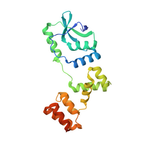
    ParB-like CTPases mediate the segregation of bacterial chromosomes and low-copy number plasmids. They act as DNA-sliding clamps that are loaded at parS motifs in the centromere of target DNA molecules and spread laterally to form large nucleoprotein complexes serving as docking points for the DNA segregation machinery. Here, we solve crystal structures of ParB in the pre- and post-hydrolysis state and illuminate the catalytic mechanism of nucleotide hydrolysis. Moreover, we identify conformational changes that underlie the CTP- and parS-dependent closure of ParB clamps. The study of CTPase-deficient ParB variants reveals that CTP hydrolysis serves to limit the sliding time of ParB clamps and thus drives the establishment of a well-defined ParB diffusion gradient across the centromere whose dynamics are critical for DNA segregation. These findings clarify the role of the ParB CTPase cycle in partition complex assembly and function and thus advance our understanding of this prototypic CTP-dependent molecular switch.


  • Organizational Affiliation

    Department of Biology, University of Marburg, 35043 Marburg, Germany; Max Planck Institute for Terrestrial Microbiology, 35043 Marburg, Germany.


Macromolecules
Find similar proteins by:  (by identity cutoff)  |  3D Structure
Entity ID: 1
MoleculeChains Sequence LengthOrganismDetailsImage
ParB family protein
A, B
213Myxococcus xanthus DK 1622Mutation(s): 0 
Gene Names: MXAN_7476
UniProt
Find proteins for Q1CVJ4 (Myxococcus xanthus (strain DK1622))
Explore Q1CVJ4 
Go to UniProtKB:  Q1CVJ4
Entity Groups  
Sequence Clusters30% Identity50% Identity70% Identity90% Identity95% Identity100% Identity
UniProt GroupQ1CVJ4
Sequence Annotations
Expand
  • Reference Sequence
Small Molecules
Ligands 5 Unique
IDChains Name / Formula / InChI Key2D Diagram3D Interactions
CDP (Subject of Investigation/LOI)
Query on CDP

Download Ideal Coordinates CCD File 
F [auth A],
K [auth B]
CYTIDINE-5'-DIPHOSPHATE
C9 H15 N3 O11 P2
ZWIADYZPOWUWEW-XVFCMESISA-N
TS6 (Subject of Investigation/LOI)
Query on TS6

Download Ideal Coordinates CCD File 
E [auth A],
J [auth B]
Monothiophosphate
H3 O3 P S
RYYWUUFWQRZTIU-UHFFFAOYSA-N
PEG
Query on PEG

Download Ideal Coordinates CCD File 
D [auth A],
H [auth B],
I [auth B]
DI(HYDROXYETHYL)ETHER
C4 H10 O3
MTHSVFCYNBDYFN-UHFFFAOYSA-N
GOL
Query on GOL

Download Ideal Coordinates CCD File 
C [auth A]GLYCEROL
C3 H8 O3
PEDCQBHIVMGVHV-UHFFFAOYSA-N
MG (Subject of Investigation/LOI)
Query on MG

Download Ideal Coordinates CCD File 
G [auth A],
L [auth B]
MAGNESIUM ION
Mg
JLVVSXFLKOJNIY-UHFFFAOYSA-N
Experimental Data & Validation

Experimental Data

  • Method: X-RAY DIFFRACTION
  • Resolution: 1.90 Å
  • R-Value Free: 0.237 
  • R-Value Work: 0.203 
  • R-Value Observed: 0.205 
  • Space Group: P 21 21 21
Unit Cell:
Length ( Å )Angle ( ˚ )
a = 44.243α = 90
b = 80.706β = 90
c = 143.38γ = 90
Software Package:
Software NamePurpose
PHENIXrefinement
XDSdata reduction
XSCALEdata scaling
PHASERphasing

Structure Validation

View Full Validation Report


Ligand Structure Quality Assessment 


Entry History 

Deposition Data

Revision History  (Full details and data files)

  • Version 1.0: 2021-09-08
    Type: Initial release
  • Version 1.1: 2021-10-13
    Changes: Data collection, Database references
  • Version 1.2: 2021-10-20
    Changes: Data collection, Database references
  • Version 1.3: 2024-01-31
    Changes: Data collection, Database references, Refinement description