A prostate-specific membrane antigen activated molecular rotor for real-time fluorescence imaging.
Zhang, J., Rakhimbekova, A., Duan, X., Yin, Q., Foss, C.A., Fan, Y., Xu, Y., Li, X., Cai, X., Kutil, Z., Wang, P., Yang, Z., Zhang, N., Pomper, M.G., Wang, Y., Barinka, C., Yang, X.(2021) Nat Commun 12: 5460-5460
- PubMed: 34526506 Search on PubMedSearch on PubMed Central
- DOI: https://doi.org/10.1038/s41467-021-25746-6
- Primary Citation Related Structures:
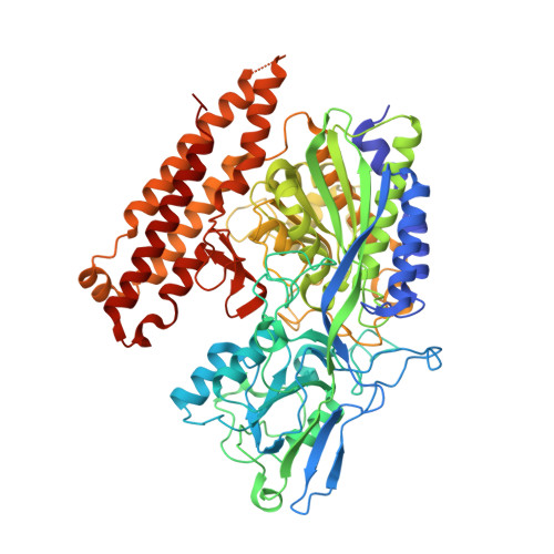
7BFZ - PubMed Abstract:
Surgery is an efficient way to treat localized prostate cancer (PCa), however, it is challenging to demarcate rapidly and accurately the tumor boundary intraoperatively, as existing tumor detection methods are seldom performed in real-time. To overcome those limitations, we develop a fluorescent molecular rotor that specifically targets the prostate-specific membrane antigen (PSMA), an established marker for PCa. The probes have picomolar affinity (IC 50 = 63-118 pM) for PSMA and generate virtually instantaneous onset of robust fluorescent signal proportional to the concentration of the PSMA-probe complex. In vitro and ex vivo experiments using PCa cell lines and clinical samples, respectively, indicate the utility of the probe for biomedical applications, including real-time monitoring of endocytosis and tumor staging. Experiments performed in a PCa xenograft model reveal suitability of the probe for imaging applications in vivo.
- Department of Nuclear Medicine, Peking University First Hospital, 100034, Beijing, China.
Organizational Affiliation: