7BBJ | pdb_00007bbj

CD73 in complex with the humanized antagonistic antibody mAb19


Experimental Data Snapshot

  • Method: X-RAY DIFFRACTION
  • Resolution: 2.72 Å
  • R-Value Free: 
    0.234 (Depositor), 0.243 (DCC) 
  • R-Value Work: 
    0.208 (Depositor), 0.216 (DCC) 
  • R-Value Observed: 
    0.210 (Depositor) 

Starting Model: experimental
View more details

wwPDB Validation 3D Report Full Report

Validation slider image for 7BBJ

This is version 1.4 of the entry. See complete history

Literature

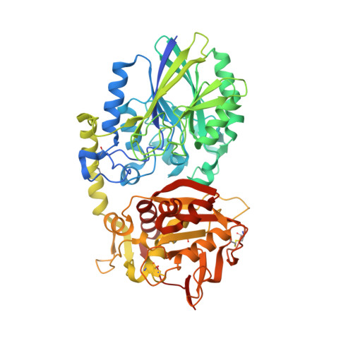
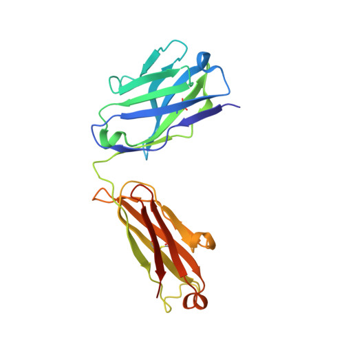
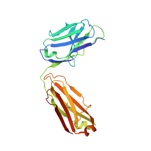
A Novel Antagonistic CD73 Antibody for Inhibition of the Immunosuppressive Adenosine Pathway.

Wurm, M.Schaaf, O.Reutner, K.Ganesan, R.Mostbock, S.Pelster, C.Bottcher, J.de Andrade Pereira, B.Taubert, C.Alt, I.Serna, G.Auguste, A.Stadermann, K.B.Delic, D.Han, F.Capdevila, J.Nuciforo, P.G.Kroe-Barrett, R.Adam, P.J.Vogt, A.B.Hofmann, I.

(2021) Mol Cancer Ther 20: 2250-2261

  • DOI: https://doi.org/10.1158/1535-7163.MCT-21-0107
  • Primary Citation Related Structures: 
    7BBJ

  • PubMed Abstract: 

    Despite some impressive clinical results with immune checkpoint inhibitors, the majority of patients with cancer do not respond to these agents, in part due to immunosuppressive mechanisms in the tumor microenvironment. High levels of adenosine in tumors can suppress immune cell function, and strategies to target the pathway involved in its production have emerged. CD73 is a key enzyme involved in adenosine production. This led us to identify a novel humanized antagonistic CD73 antibody, mAb19, with distinct binding properties. mAb19 potently inhibits the enzymatic activity of CD73 in vitro , resulting in an inhibition of adenosine formation and enhanced T-cell activation. We then investigated the therapeutic potential of combining CD73 antagonism with other immune modulatory and chemotherapeutic agents. Combination of mAb19 with a PD-1 inhibitor increased T-cell activation in vitro Interestingly, this effect could be further enhanced with an agonist of the adenosine receptor ADORA3. Adenosine levels were found to be elevated upon doxorubicin treatment in vivo , which could be blocked by CD73 inhibition. Combining CD73 antagonism with doxorubicin resulted in superior responses in vivo Furthermore, a retrospective analysis of rectal cancer patient samples demonstrated an upregulation of the adenosine pathway upon chemoradiation, providing further rationale for combining CD73 inhibition with chemotherapeutic agents.This study demonstrates the ability of a novel CD73 antibody to enhance T-cell function through the potent suppression of adenosine levels. In addition, the data highlight combination opportunities with standard of care therapies as well as with an ADORA3 receptor agonist to treat patients with solid tumors.


  • Organizational Affiliation
    • Boehringer Ingelheim RCV, GmbH & Co KG, Cancer Pharmacology and Disease Positioning, Vienna, Austria.

Macromolecule Content 

  • Total Structure Weight: 216.42 kDa 
  • Atom Count: 14,874 
  • Modeled Residue Count: 1,892 
  • Deposited Residue Count: 1,964 
  • Unique protein chains: 3

Macromolecules

Find similar proteins by:|  3D Structure
Entity ID: 1
MoleculeChains  Sequence LengthOrganismDetailsImage
5'-nucleotidase
A, B
546Homo sapiensMutation(s): 7 
Gene Names: NT5ENT5NTE
EC: 3.1.3.5 (PDB Primary Data), 3.1.3.99 (UniProt), 3.1.3.89 (UniProt), 3.1.3.35 (UniProt), 3.1.3.91 (UniProt)
UniProt & NIH Common Fund Data Resources
Find proteins for P21589 (Homo sapiens)
Explore P21589 
Go to UniProtKB:  P21589
PHAROS:  P21589
GTEx:  ENSG00000135318 
Entity Groups
Sequence Clusters30% Identity50% Identity70% Identity90% Identity95% Identity100% Identity
UniProt GroupP21589
Sequence Annotations
Expand
Reference Sequence
Find similar proteins by:|  3D Structure
Entity ID: 2
MoleculeChains  Sequence LengthOrganismDetailsImage
heavy chain mAb19C [auth H],
E [auth M]
222synthetic constructMutation(s): 0 
Find similar proteins by:|  3D Structure
Entity ID: 3
MoleculeChains  Sequence LengthOrganismDetailsImage
light chain mAB19D [auth L],
F [auth N]
214synthetic constructMutation(s): 0 

Experimental Data & Validation

Experimental Data

  • Method: X-RAY DIFFRACTION
  • Resolution: 2.72 Å
  • R-Value Free:  0.234 (Depositor), 0.243 (DCC) 
  • R-Value Work:  0.208 (Depositor), 0.216 (DCC) 
  • R-Value Observed: 0.210 (Depositor) 
Space Group: P 1 21 1
Unit Cell:
Length ( Å )Angle ( ˚ )
a = 133.35α = 90
b = 67.74β = 100.13
c = 150.05γ = 90
Software Package:
Software NamePurpose
BUSTERrefinement
XDSdata reduction
STARANISOdata scaling
MOLREPphasing

Structure Validation

View Full Validation Report


Entry History 

& Funding Information

Deposition Data


Funding OrganizationLocationGrant Number
Austrian Research Promotion AgencyAustraliaFFG; 852068

Revision History  (Full details and data files)

  • Version 1.0: 2021-12-29
    Type: Initial release
  • Version 1.1: 2022-01-05
    Changes: Database references
  • Version 1.2: 2022-10-26
    Changes: Source and taxonomy
  • Version 1.3: 2024-01-31
    Changes: Data collection, Refinement description
  • Version 1.4: 2024-11-06
    Changes: Structure summary