7BBI

Joint X-ray/neutron room temperature structure of H/D-exchanged PLL lectin


Experimental Data Snapshot

  • Method: X-RAY DIFFRACTION
  • Resolution: 1.70 Å
  • R-Value Free: 0.156 
  • R-Value Work: 0.135 
  • R-Value Observed: 0.136 

  • Method: NEUTRON DIFFRACTION
  • Resolution: 2.20 Å
  • R-Value Free: 0.240 
  • R-Value Work: 0.215 
  • R-Value Observed: 0.216 

wwPDB Validation   3D Report Full Report


This is version 1.2 of the entry. See complete history


Literature

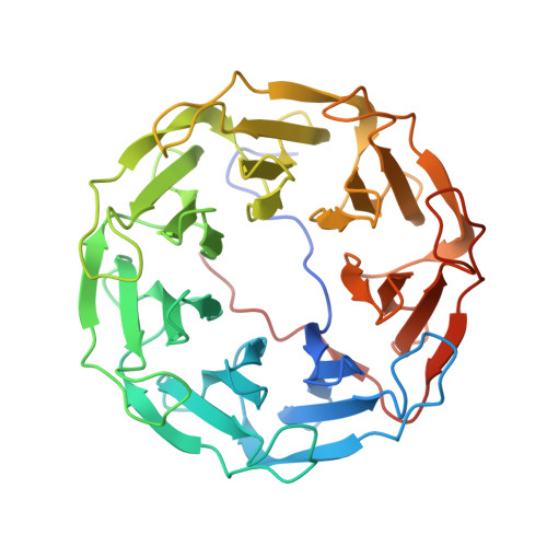
Visualization of hydrogen atoms in a perdeuterated lectin-fucose complex reveals key details of protein-carbohydrate interactions.

Gajdos, L.Blakeley, M.P.Kumar, A.Wimmerova, M.Haertlein, M.Forsyth, V.T.Imberty, A.Devos, J.M.

(2021) Structure 29: 1003-1013.e4

  • DOI: https://doi.org/10.1016/j.str.2021.03.003
  • Primary Citation of Related Structures:  
    7B7C, 7B7E, 7B7F, 7BB4, 7BBC, 7BBI

  • PubMed Abstract: 

    Carbohydrate-binding proteins from pathogenic bacteria and fungi have been shown to be implicated in various pathological processes, where they interact with glycans present on the surface of the host cells. These interactions are part of the initial processes of infection of the host and are very important to study at the atomic level. Here, we report the room temperature neutron structures of PLL lectin from Photorhabdus laumondii in its apo form and in complex with deuterated L-fucose, which is, to our knowledge, the first neutron structure of a carbohydrate-binding protein in complex with a fully deuterated carbohydrate ligand. A detailed structural analysis of the lectin-carbohydrate interactions provides information on the hydrogen bond network, the role of water molecules, and the extent of the CH-π stacking interactions between fucose and the aromatic amino acids in the binding site.


  • Organizational Affiliation

    Life Sciences Group, Institut Laue-Langevin, 38000 Grenoble, France; Partnership for Structural Biology (PSB), 38000 Grenoble, France; Université Grenoble Alpes, CNRS, CERMAV, 38000 Grenoble, France.


Macromolecules
Find similar proteins by:  (by identity cutoff)  |  3D Structure
Entity ID: 1
MoleculeChains Sequence LengthOrganismDetailsImage
PLL lectin381Photorhabdus laumondiiMutation(s): 0 
Gene Names: CKY10_20885
UniProt
Find proteins for Q7N8J0 (Photorhabdus laumondii subsp. laumondii (strain DSM 15139 / CIP 105565 / TT01))
Explore Q7N8J0 
Go to UniProtKB:  Q7N8J0
Entity Groups  
Sequence Clusters30% Identity50% Identity70% Identity90% Identity95% Identity100% Identity
UniProt GroupQ7N8J0
Sequence Annotations
Expand
  • Reference Sequence
Experimental Data & Validation

Experimental Data

  • Method: X-RAY DIFFRACTION
  • Resolution: 1.70 Å
  • R-Value Free: 0.156 
  • R-Value Work: 0.135 
  • R-Value Observed: 0.136 
  • Space Group: I 2 2 2
  • Method: NEUTRON DIFFRACTION
  • Resolution: 2.20 Å
  • R-Value Free: 0.240 
  • R-Value Work: 0.215 
  • R-Value Observed: 0.216 
  • Space Group: I 2 2 2
Unit Cell:
Length ( Å )Angle ( ˚ )
a = 72.68α = 90
b = 89.34β = 90
c = 159.38γ = 90
Software Package:
Software NamePurpose
PHENIXrefinement
XDSdata reduction
XSCALEdata scaling
SCALAdata scaling
PHENIXphasing

Structure Validation

View Full Validation Report


Entry History & Funding Information

Deposition Data


Funding OrganizationLocationGrant Number
Other privateFrance--

Revision History  (Full details and data files)

  • Version 1.0: 2021-03-17
    Type: Initial release
  • Version 1.1: 2021-04-14
    Changes: Database references
  • Version 1.2: 2021-09-15
    Changes: Database references