7BB5 | pdb_00007bb5

Crystal structure of anti-CRISPR protein AcrIF9


Experimental Data Snapshot

  • Method: X-RAY DIFFRACTION
  • Resolution: 2.30 Å
  • R-Value Free: 
    0.223 (Depositor), 0.241 (DCC) 
  • R-Value Work: 
    0.183 (Depositor) 
  • R-Value Observed: 
    0.187 (Depositor) 

Starting Model: experimental
View more details

wwPDB Validation 3D Report Full Report

Validation slider image for 7BB5

This is version 1.2 of the entry. See complete history

Literature

Disarming of type I-F CRISPR-Cas surveillance complex by anti-CRISPR proteins AcrIF6 and AcrIF9.

Kupcinskaite, E.Tutkus, M.Kopustas, A.Asmontas, S.Jankunec, M.Zaremba, M.Tamulaitiene, G.Sinkunas, T.

(2022) Sci Rep 12: 15548-15548

  • DOI: https://doi.org/10.1038/s41598-022-19797-y
  • Primary Citation Related Structures: 
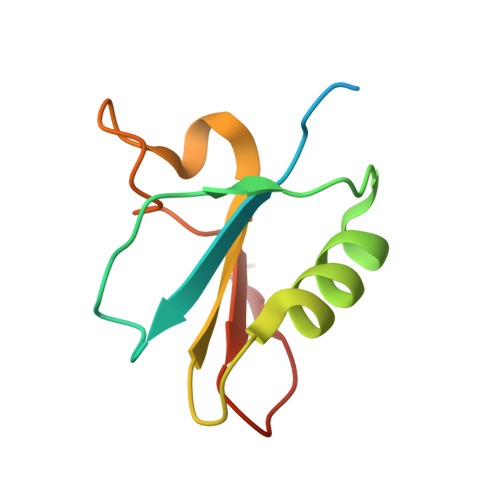
    7BB5

  • PubMed Abstract: 

    CRISPR-Cas systems are prokaryotic adaptive immune systems that protect against phages and other invading nucleic acids. The evolutionary arms race between prokaryotes and phages gave rise to phage anti-CRISPR (Acr) proteins that act as a counter defence against CRISPR-Cas systems by inhibiting the effector complex. Here, we used a combination of bulk biochemical experiments, X-ray crystallography and single-molecule techniques to explore the inhibitory activity of AcrIF6 and AcrIF9 proteins against the type I-F CRISPR-Cas system from Aggregatibacter actinomycetemcomitans (Aa). We showed that AcrIF6 and AcrIF9 proteins hinder Aa-Cascade complex binding to target DNA. We solved a crystal structure of Aa1-AcrIF9 protein, which differ from other known AcrIF9 proteins by an additional structurally important loop presumably involved in the interaction with Cascade. We revealed that AcrIF9 association with Aa-Cascade promotes its binding to off-target DNA sites, which facilitates inhibition of CRISPR-Cas protection.


  • Organizational Affiliation
    • Institute of Biotechnology, Life Sciences Center, Vilnius University, Sauletekio Ave. 7, 10257, Vilnius, Lithuania.

Macromolecule Content 

  • Total Structure Weight: 10.33 kDa 
  • Atom Count: 666 
  • Modeled Residue Count: 76 
  • Deposited Residue Count: 91 
  • Unique protein chains: 1

Macromolecules

Find similar proteins by:|  3D Structure
Entity ID: 1
MoleculeChains  Sequence LengthOrganismDetailsImage
AcrIF991Aggregatibacter actinomycetemcomitans serotype e str. SC1083Mutation(s): 0 
Gene Names: SC1083_0445
UniProt
Find proteins for G4A6K5 (Aggregatibacter actinomycetemcomitans serotype e str. SC1083)
Explore G4A6K5 
Go to UniProtKB:  G4A6K5
Entity Groups
Sequence Clusters30% Identity50% Identity70% Identity90% Identity95% Identity100% Identity
UniProt GroupG4A6K5
Sequence Annotations
Expand
Reference Sequence

Experimental Data & Validation

Experimental Data

  • Method: X-RAY DIFFRACTION
  • Resolution: 2.30 Å
  • R-Value Free:  0.223 (Depositor), 0.241 (DCC) 
  • R-Value Work:  0.183 (Depositor) 
  • R-Value Observed: 0.187 (Depositor) 
Space Group: I 41 2 2
Unit Cell:
Length ( Å )Angle ( ˚ )
a = 77.285α = 90
b = 77.285β = 90
c = 75.663γ = 90
Software Package:
Software NamePurpose
XDSdata reduction
SCALAdata scaling
MOLREPphasing
PHENIXrefinement
PDB_EXTRACTdata extraction

Structure Validation

View Full Validation Report


Entry History 

& Funding Information

Deposition Data


Funding OrganizationLocationGrant Number
Research Council of LithuaniaLithuaniaS-MIP-17-47

Revision History  (Full details and data files)

  • Version 1.0: 2021-11-24
    Type: Initial release
  • Version 1.1: 2022-12-07
    Changes: Database references
  • Version 1.2: 2024-05-01
    Changes: Data collection, Refinement description