7B9P

Structure of Ribonucleotide reductase from Rhodobacter sphaeroides


Experimental Data Snapshot

  • Method: X-RAY DIFFRACTION
  • Resolution: 2.65 Å
  • R-Value Free: 0.272 
  • R-Value Work: 0.230 
  • R-Value Observed: 0.231 

wwPDB Validation   3D Report Full Report


Ligand Structure Quality Assessment 


This is version 1.2 of the entry. See complete history


Literature

HUG Domain Is Responsible for Active Dimer Stabilization in an NrdJd Ribonucleotide Reductase.

Fietze, T.Wilk, P.Kabinger, F.Anoosheh, S.Hofer, A.Lundin, D.Feiler, C.G.Weiss, M.S.Loderer, C.

(2022) Biochemistry 61: 1633-1641

  • DOI: https://doi.org/10.1021/acs.biochem.2c00173
  • Primary Citation of Related Structures:  
    7B9P, 7B9Q

  • PubMed Abstract: 

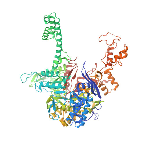
    Ribonucleotide reductases (RNRs) catalyze the reduction of ribonucleotides to the corresponding deoxyribonucleotides. The catalytic activity of most RNRs depends on the formation of a dimer of the catalytic subunits. The active site is located at the interface, and part of the substrate binding site and regulatory mechanisms work across the subunit in the dimer. In this study, we describe and characterize a novel domain responsible for forming the catalytic dimer in several class II RNRs. The 3D structure of the class II RNR from Rhodobacter sphaeroides reveals a so far undescribed α-helical domain in the dimer interface, which is embracing the other subunit. Genetic removal of this HUG domain leads to a severe reduction of activity paired with reduced dimerization capability. In comparison with other described RNRs, the enzyme with this domain is less dependent on the presence of nucleotides to act as allosteric effectors in the formation of dimers. The HUG domain appears to serve as an interlock to keep the dimer intact and functional even at low enzyme and/or effector concentrations.


  • Organizational Affiliation

    Chair of Molecular Biotechnology, Technische Universität Dresden, Dresden 01217, Germany.


Macromolecules
Find similar proteins by:  (by identity cutoff)  |  3D Structure
Entity ID: 1
MoleculeChains Sequence LengthOrganismDetailsImage
Vitamin B12-dependent ribonucleotide reductase925Cereibacter sphaeroides 2.4.1Mutation(s): 0 
Gene Names: nrdRSP_2495
EC: 1.17.4.1
UniProt
Find proteins for Q3J3H6 (Cereibacter sphaeroides (strain ATCC 17023 / DSM 158 / JCM 6121 / CCUG 31486 / LMG 2827 / NBRC 12203 / NCIMB 8253 / ATH 2.4.1.))
Explore Q3J3H6 
Go to UniProtKB:  Q3J3H6
Entity Groups  
Sequence Clusters30% Identity50% Identity70% Identity90% Identity95% Identity100% Identity
UniProt GroupQ3J3H6
Sequence Annotations
Expand
  • Reference Sequence
Small Molecules
Ligands 2 Unique
IDChains Name / Formula / InChI Key2D Diagram3D Interactions
DTP
Query on DTP

Download Ideal Coordinates CCD File 
B [auth A]2'-DEOXYADENOSINE 5'-TRIPHOSPHATE
C10 H16 N5 O12 P3
SUYVUBYJARFZHO-RRKCRQDMSA-N
MG
Query on MG

Download Ideal Coordinates CCD File 
C [auth A]MAGNESIUM ION
Mg
JLVVSXFLKOJNIY-UHFFFAOYSA-N
Experimental Data & Validation

Experimental Data

  • Method: X-RAY DIFFRACTION
  • Resolution: 2.65 Å
  • R-Value Free: 0.272 
  • R-Value Work: 0.230 
  • R-Value Observed: 0.231 
  • Space Group: P 65 2 2
Unit Cell:
Length ( Å )Angle ( ˚ )
a = 140.748α = 90
b = 140.748β = 90
c = 364.277γ = 120
Software Package:
Software NamePurpose
PHENIXrefinement
PDB_EXTRACTdata extraction
XDSdata reduction

Structure Validation

View Full Validation Report


Ligand Structure Quality Assessment 


Entry History 

Deposition Data

Revision History  (Full details and data files)

  • Version 1.0: 2022-01-12
    Type: Initial release
  • Version 1.1: 2022-08-03
    Changes: Database references
  • Version 1.2: 2022-08-10
    Changes: Database references