


Crystal structure of MurE from E.coli
Koekemoer, L., Steindel, M., Fairhead, M., Talon, R., Douangamath, A., Arrowsmith, C.H., Edwards, A.M., Bountra, C., von Delft, F., Krojer, T.To be published.
Experimental Data Snapshot
Starting Model: experimental
View more details
Entity ID: 1 | |||||
|---|---|---|---|---|---|
| Molecule | Chains | Sequence Length | Organism | Details | Image |
| UDP-N-acetylmuramoyl-L-alanyl-D-glutamate-2,6-diaminopimelate ligase | 496 | Escherichia coli K-12 | Mutation(s): 0 Gene Names: murE, b0085, JW0083 EC: 6.3.2.13 | 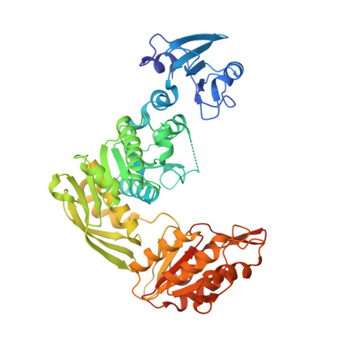 | |
UniProt | |||||
Entity Groups | |||||
| Sequence Clusters | 30% Identity50% Identity70% Identity90% Identity95% Identity100% Identity | ||||
| UniProt Group | P22188 | ||||
Sequence AnnotationsExpand | |||||
Reference Sequence | |||||
| Ligands 3 Unique | |||||
|---|---|---|---|---|---|
| ID | Chains | Name / Formula / InChI Key | 2D Diagram | 3D Interactions | |
| O3D (Subject of Investigation/LOI) Download:Ideal Coordinates CCD File | C [auth A], D [auth A], F [auth B] | 4-[(furan-2-yl)methyl]-1lambda~6~,4-thiazinane-1,1-dione C9 H13 N O3 S AEOGPIJVSQTAII-UHFFFAOYSA-N |  | ||
| DMS Download:Ideal Coordinates CCD File | E [auth A], G [auth B] | DIMETHYL SULFOXIDE C2 H6 O S IAZDPXIOMUYVGZ-UHFFFAOYSA-N |  | ||
| IPA Download:Ideal Coordinates CCD File | H [auth B], I [auth B] | ISOPROPYL ALCOHOL C3 H8 O KFZMGEQAYNKOFK-UHFFFAOYSA-N |  | ||
| Length ( Å ) | Angle ( ˚ ) |
|---|---|
| a = 58.249 | α = 97.27 |
| b = 58.382 | β = 91.53 |
| c = 74.027 | γ = 105.61 |
| Software Name | Purpose |
|---|---|
| REFMAC | refinement |
| xia2 | data reduction |
| xia2 | data scaling |
| PHASER | phasing |


