7B5J

Anti-CRISPR associated (Aca) protein, Aca2


Experimental Data Snapshot

  • Method: X-RAY DIFFRACTION
  • Resolution: 1.34 Å
  • R-Value Free: 0.176 
  • R-Value Work: 0.148 
  • R-Value Observed: 0.149 

wwPDB Validation   3D Report Full Report


This is version 1.2 of the entry. See complete history


Literature

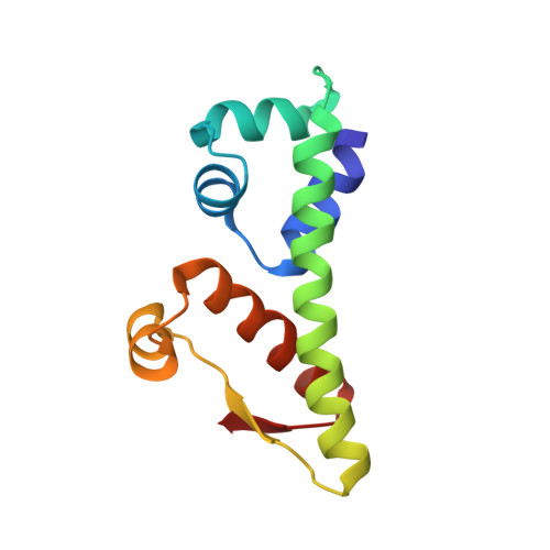
Crystal structure of the anti-CRISPR repressor Aca2.

Usher, B.Birkholz, N.Beck, I.N.Fagerlund, R.D.Jackson, S.A.Fineran, P.C.Blower, T.R.

(2021) J Struct Biol 213: 107752-107752

  • DOI: https://doi.org/10.1016/j.jsb.2021.107752
  • Primary Citation of Related Structures:  
    7B5J, 7ZGE

  • PubMed Abstract: 

    Bacteria use adaptive CRISPR-Cas immune mechanisms to protect from invasion by bacteriophages and other mobile genetic elements. In response, bacteriophages and mobile genetic elements have co-evolved anti-CRISPR proteins to inhibit the bacterial defense. We and others have previously shown that anti-CRISPR associated (Aca) proteins can regulate this anti-CRISPR counter-attack. Here, we report the first structure of an Aca protein, the Aca2 DNA-binding transcriptional autorepressor from Pectobacterium carotovorum bacteriophage ZF40, determined to 1.34 Å. Aca2 presents a conserved N-terminal helix-turn-helix DNA-binding domain and a previously uncharacterized C-terminal dimerization domain. Dimerization positions the Aca2 recognition helices for insertion into the major grooves of target DNA, supporting its role in regulating anti-CRISPRs. Furthermore, database comparisons identified uncharacterized Aca2 structural homologs in pathogenic bacteria, suggesting that Aca2 represents the first characterized member of a more widespread family of transcriptional regulators.


  • Organizational Affiliation

    Department of Biosciences, Durham University, Stockton Road, Durham DH1 3LE, UK.


Macromolecules
Find similar proteins by:  (by identity cutoff)  |  3D Structure
Entity ID: 1
MoleculeChains Sequence LengthOrganismDetailsImage
Anti-CRISPR associated (Aca) protein, Aca2
A, B
115Pectobacterium phage ZF40Mutation(s): 0 
Gene Names: ZF40_0030aca2
UniProt
Find proteins for H9C180 (Pectobacterium phage ZF40)
Explore H9C180 
Go to UniProtKB:  H9C180
Entity Groups  
Sequence Clusters30% Identity50% Identity70% Identity90% Identity95% Identity100% Identity
UniProt GroupH9C180
Sequence Annotations
Expand
  • Reference Sequence
Small Molecules
Ligands 1 Unique
IDChains Name / Formula / InChI Key2D Diagram3D Interactions
GOL
Query on GOL

Download Ideal Coordinates CCD File 
C [auth A]GLYCEROL
C3 H8 O3
PEDCQBHIVMGVHV-UHFFFAOYSA-N
Experimental Data & Validation

Experimental Data

  • Method: X-RAY DIFFRACTION
  • Resolution: 1.34 Å
  • R-Value Free: 0.176 
  • R-Value Work: 0.148 
  • R-Value Observed: 0.149 
  • Space Group: P 1 21 1
Unit Cell:
Length ( Å )Angle ( ˚ )
a = 39.791α = 90
b = 67.103β = 106.331
c = 42.24γ = 90
Software Package:
Software NamePurpose
GDAdata collection
xia2data reduction
Arcimboldophasing
BUCCANEERmodel building
Cootmodel building
PHENIXrefinement
xia2data scaling

Structure Validation

View Full Validation Report


Entry History 

Deposition Data

Revision History  (Full details and data files)

  • Version 1.0: 2021-06-23
    Type: Initial release
  • Version 1.1: 2021-06-30
    Changes: Database references
  • Version 1.2: 2021-08-25
    Changes: Database references, Structure summary