7B5E

Structure of calcium-bound mTMEM16A(ac)-I551A chloride channel at 4.1 A resolution


Experimental Data Snapshot

  • Method: ELECTRON MICROSCOPY
  • Resolution: 4.10 Å
  • Aggregation State: PARTICLE 
  • Reconstruction Method: SINGLE PARTICLE 

wwPDB Validation   3D Report Full Report


This is version 1.1 of the entry. See complete history


Literature

Gating the pore of the calcium-activated chloride channel TMEM16A.

Lam, A.K.M.Rheinberger, J.Paulino, C.Dutzler, R.

(2021) Nat Commun 12: 785-785

  • DOI: https://doi.org/10.1038/s41467-020-20787-9
  • Primary Citation of Related Structures:  
    7B5C, 7B5D, 7B5E

  • PubMed Abstract: 

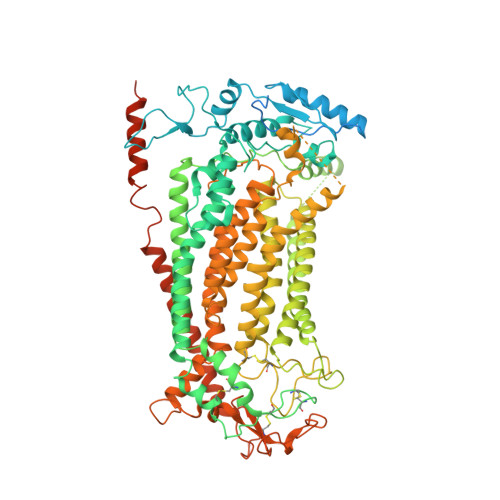
    The binding of cytoplasmic Ca 2+ to the anion-selective channel TMEM16A triggers a conformational change around its binding site that is coupled to the release of a gate at the constricted neck of an hourglass-shaped pore. By combining mutagenesis, electrophysiology, and cryo-electron microscopy, we identified three hydrophobic residues at the intracellular entrance of the neck as constituents of this gate. Mutation of each of these residues increases the potency of Ca 2+ and results in pronounced basal activity. The structure of an activating mutant shows a conformational change of an α-helix that contributes to Ca 2+ binding as a likely cause for the basal activity. Although not in physical contact, the three residues are functionally coupled to collectively contribute to the stabilization of the gate in the closed conformation of the pore, thus explaining the low open probability of the channel in the absence of Ca 2+ .


  • Organizational Affiliation

    Department of Biochemistry, University of Zurich, Winterthurerstrasse 190, CH-8057, Zurich, Switzerland. a.lam@bioc.uzh.ch.


Macromolecules
Find similar proteins by:  (by identity cutoff)  |  3D Structure
Entity ID: 1
MoleculeChains Sequence LengthOrganismDetailsImage
Anoctamin-1
A, B
960Mus musculusMutation(s): 1 
Gene Names: Ano1Tmem16a
Membrane Entity: Yes 
UniProt
Find proteins for Q8BHY3 (Mus musculus)
Explore Q8BHY3 
Go to UniProtKB:  Q8BHY3
Entity Groups  
Sequence Clusters30% Identity50% Identity70% Identity90% Identity95% Identity100% Identity
UniProt GroupQ8BHY3
Sequence Annotations
Expand
  • Reference Sequence
Experimental Data & Validation

Experimental Data

  • Method: ELECTRON MICROSCOPY
  • Resolution: 4.10 Å
  • Aggregation State: PARTICLE 
  • Reconstruction Method: SINGLE PARTICLE 
EM Software:
TaskSoftware PackageVersion
RECONSTRUCTIONRELION3.0
MODEL REFINEMENTPHENIX1.16

Structure Validation

View Full Validation Report


Entry History & Funding Information

Deposition Data


Funding OrganizationLocationGrant Number
European Research Council (ERC)Switzerland339116
Netherlands Organisation for Scientific Research (NWO)Netherlands722.017.001
Netherlands Organisation for Scientific Research (NWO)Netherlands740.018.016
University of ZurichSwitzerlandFK-18-048

Revision History  (Full details and data files)

  • Version 1.0: 2021-02-10
    Type: Initial release
  • Version 1.1: 2021-02-17
    Changes: Database references