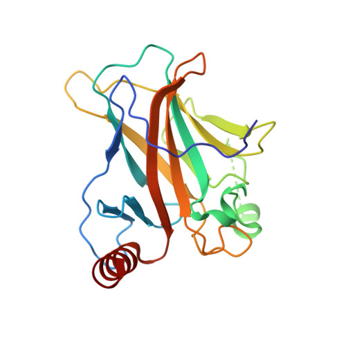
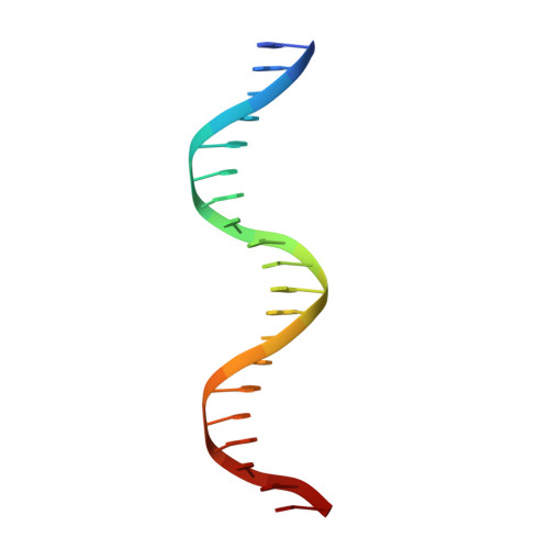
Structural basis of reactivation of oncogenic p53 mutants by a small molecule: methylene quinuclidinone (MQ).
Degtjarik, O., Golovenko, D., Diskin-Posner, Y., Abrahmsen, L., Rozenberg, H., Shakked, Z.(2021) Nat Commun 12: 7057-7057
- PubMed: 34862374 Search on PubMedSearch on PubMed Central
- DOI: https://doi.org/10.1038/s41467-021-27142-6
- Primary Citation Related Structures:
6ZNC, 7B46, 7B47, 7B48, 7B49, 7B4A, 7B4B, 7B4C, 7B4D, 7B4E, 7B4F, 7B4G, 7B4H, 7B4N - PubMed Abstract:
In response to genotoxic stress, the tumor suppressor p53 acts as a transcription factor by regulating the expression of genes critical for cancer prevention. Mutations in the gene encoding p53 are associated with cancer development. PRIMA-1 and eprenetapopt (APR-246/PRIMA-1 MET ) are small molecules that are converted into the biologically active compound, methylene quinuclidinone (MQ), shown to reactivate mutant p53 by binding covalently to cysteine residues. Here, we investigate the structural basis of mutant p53 reactivation by MQ based on a series of high-resolution crystal structures of cancer-related and wild-type p53 core domains bound to MQ in their free state and in complexes with their DNA response elements. Our data demonstrate that MQ binds to several cysteine residues located at the surface of the core domain. The structures reveal a large diversity in MQ interaction modes that stabilize p53 and its complexes with DNA, leading to a common global effect that is pertinent to the restoration of non-functional p53 proteins.
- Department of Chemical and Structural Biology, Weizmann Institute of Science, 76100, Rehovot, Israel.
Organizational Affiliation: