7B3G | pdb_00007b3g

Notum complex with ARUK3003902


Experimental Data Snapshot

  • Method: X-RAY DIFFRACTION
  • Resolution: 1.28 Å
  • R-Value Free: 
    0.215 (Depositor), 0.210 (DCC) 
  • R-Value Work: 
    0.202 (Depositor) 
  • R-Value Observed: 
    0.203 (Depositor) 

Starting Model: experimental
View more details

wwPDB Validation 3D Report Full Report

Validation slider image for 7B3G

Ligand Structure Quality Assessment 


This is version 1.3 of the entry. See complete history

Literature

Virtual Screening Directly Identifies New Fragment-Sized Inhibitors of Carboxylesterase Notum with Nanomolar Activity.

Steadman, D.Atkinson, B.N.Zhao, Y.Willis, N.J.Frew, S.Monaghan, A.Patel, C.Armstrong, E.Costelloe, K.Magno, L.Bictash, M.Jones, E.Y.Fish, P.V.Svensson, F.

(2022) J Med Chem 65: 562-578

  • DOI: https://doi.org/10.1021/acs.jmedchem.1c01735
  • Primary Citation Related Structures: 
    7B3G, 7B3H, 7B3I, 7B3P, 7B3X, 7B45, 7B50

  • PubMed Abstract: 

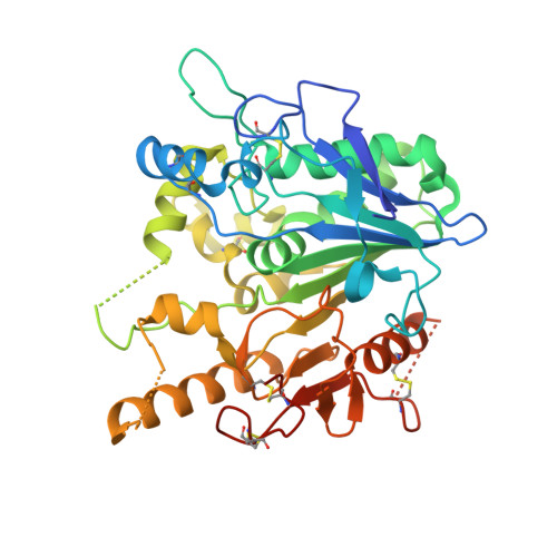
    Notum is a negative regulator of Wnt signaling acting through the hydrolysis of a palmitoleoylate ester, which is required for Wnt activity. Inhibitors of Notum could be of use in diseases where dysfunctional Notum activity is an underlying cause. A docking-based virtual screen (VS) of a large commercial library was used to shortlist 952 compounds for experimental validation as inhibitors of Notum. The VS was successful with 31 compounds having an IC 50 < 500 nM. A critical selection process was then applied with two clusters and two singletons ( 1 - 4d ) selected for hit validation. Optimization of 4d guided by structural biology identified potent inhibitors of Notum activity that restored Wnt/β-catenin signaling in cell-based models. The [1,2,4]triazolo[4,3- b ]pyradizin-3(2 H )-one series 4 represent a new chemical class of Notum inhibitors and the first to be discovered by a VS campaign. These results demonstrate the value of VS with well-designed docking models based on X-ray structures.


  • Organizational Affiliation
    • Alzheimer's Research UK UCL Drug Discovery Institute, University College London, The Cruciform Building, Gower Street, LondonWC1E 6BT, U.K.

Macromolecule Content 

  • Total Structure Weight: 45.1 kDa 
  • Atom Count: 2,942 
  • Modeled Residue Count: 344 
  • Deposited Residue Count: 383 
  • Unique protein chains: 1

Macromolecules

Find similar proteins by:|  3D Structure
Entity ID: 1
MoleculeChains  Sequence LengthOrganismDetailsImage
Palmitoleoyl-protein carboxylesterase NOTUM383Homo sapiensMutation(s): 1 
Gene Names: NOTUMOK/SW-CL.30
EC: 3.1.1.98
UniProt & NIH Common Fund Data Resources
Find proteins for Q6P988 (Homo sapiens)
Explore Q6P988 
Go to UniProtKB:  Q6P988
PHAROS:  Q6P988
GTEx:  ENSG00000185269 
Entity Groups
Sequence Clusters30% Identity50% Identity70% Identity90% Identity95% Identity100% Identity
UniProt GroupQ6P988
Glycosylation
Glycosylation Sites: 1Go to GlyGen: Q6P988-1
Sequence Annotations
Expand
Reference Sequence

Small Molecules

Ligands 5 Unique
IDChains Name / Formula / InChI Key2D Diagram3D Interactions
SSQ
(Subject of Investigation/LOI)

Query on SSQ


Download:Ideal Coordinates CCD File
I [auth A]6-((2-chlorophenyl)thio)-[1,2,4]triazolo[4,3-b]pyridazin-3(2H)-one
C11 H7 Cl N4 O S
IOETYCRGZYHINY-UHFFFAOYSA-N
NAG

Query on NAG


Download:Ideal Coordinates CCD File
B [auth A]2-acetamido-2-deoxy-beta-D-glucopyranose
C8 H15 N O6
OVRNDRQMDRJTHS-FMDGEEDCSA-N
SO4

Query on SO4


Download:Ideal Coordinates CCD File
C [auth A]
D [auth A]
E [auth A]
L [auth A]
M [auth A]
C [auth A],
D [auth A],
E [auth A],
L [auth A],
M [auth A],
N [auth A],
O [auth A]
SULFATE ION
O4 S
QAOWNCQODCNURD-UHFFFAOYSA-L
DMS

Query on DMS


Download:Ideal Coordinates CCD File
F [auth A],
J [auth A],
K [auth A]
DIMETHYL SULFOXIDE
C2 H6 O S
IAZDPXIOMUYVGZ-UHFFFAOYSA-N
EDO

Query on EDO


Download:Ideal Coordinates CCD File
G [auth A],
H [auth A]
1,2-ETHANEDIOL
C2 H6 O2
LYCAIKOWRPUZTN-UHFFFAOYSA-N
Binding Affinity Annotations 
IDSourceBinding Affinity
SSQ BindingDB:  7B3G IC50: 30 (nM) from 1 assay(s)
EC50: 720 (nM) from 1 assay(s)

Experimental Data & Validation

Experimental Data

  • Method: X-RAY DIFFRACTION
  • Resolution: 1.28 Å
  • R-Value Free:  0.215 (Depositor), 0.210 (DCC) 
  • R-Value Work:  0.202 (Depositor) 
  • R-Value Observed: 0.203 (Depositor) 
Space Group: P 21 21 21
Unit Cell:
Length ( Å )Angle ( ˚ )
a = 59.42α = 90
b = 72.54β = 90
c = 78.065γ = 90
Software Package:
Software NamePurpose
xia2data scaling
PHENIXrefinement
PDB_EXTRACTdata extraction
xia2data reduction
MOLREPphasing

Structure Validation

View Full Validation Report


Ligand Structure Quality Assessment 


Entry History 

Deposition Data

Revision History  (Full details and data files)

  • Version 1.0: 2021-12-08
    Type: Initial release
  • Version 1.1: 2023-06-21
    Changes: Database references
  • Version 1.2: 2024-02-07
    Changes: Data collection, Refinement description
  • Version 1.3: 2024-10-09
    Changes: Structure summary