7B16 | pdb_00007b16

TRPC4 in complex with inhibitor GFB-9289


Experimental Data Snapshot

  • Method: ELECTRON MICROSCOPY
  • Resolution: 3.15 Å
  • Aggregation State: PARTICLE 
  • Reconstruction Method: SINGLE PARTICLE 

Starting Model: experimental
View more details

wwPDB Validation 3D Report Full Report

Validation slider image for 7B16

This is version 1.1 of the entry. See complete history

Literature

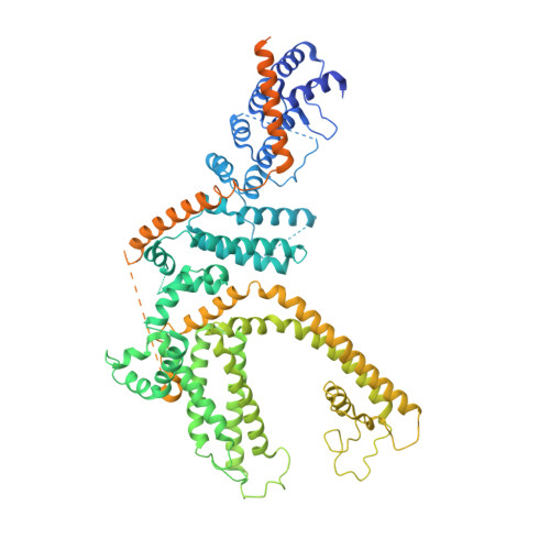
Structural basis of TRPC4 regulation by calmodulin and pharmacological agents.

Vinayagam, D.Quentin, D.Yu-Strzelczyk, J.Sitsel, O.Merino, F.Stabrin, M.Hofnagel, O.Yu, M.Ledeboer, M.W.Nagel, G.Malojcic, G.Raunser, S.

(2020) Elife 9

  • DOI: https://doi.org/10.7554/eLife.60603
  • Primary Citation Related Structures: 
    7B05, 7B0J, 7B0S, 7B16, 7B1G

  • PubMed Abstract: 

    Canonical transient receptor potential channels (TRPC) are involved in receptor-operated and/or store-operated Ca 2+ signaling. Inhibition of TRPCs by small molecules was shown to be promising in treating renal diseases. In cells, the channels are regulated by calmodulin (CaM). Molecular details of both CaM and drug binding have remained elusive so far. Here, we report structures of TRPC4 in complex with three pyridazinone-based inhibitors and CaM. The structures reveal that all the inhibitors bind to the same cavity of the voltage-sensing-like domain and allow us to describe how structural changes from the ligand-binding site can be transmitted to the central ion-conducting pore of TRPC4. CaM binds to the rib helix of TRPC4, which results in the ordering of a previously disordered region, fixing the channel in its closed conformation. This represents a novel CaM-induced regulatory mechanism of canonical TRP channels.


  • Organizational Affiliation
    • Department of Structural Biochemistry, Max Planck Institute of Molecular Physiology, Dortmund, Germany.

Macromolecule Content 

  • Total Structure Weight: 424.82 kDa 
  • Atom Count: 21,152 
  • Modeled Residue Count: 2,572 
  • Deposited Residue Count: 3,660 
  • Unique protein chains: 1

Macromolecules

Find similar proteins by:|  3D Structure
Entity ID: 1
MoleculeChains  Sequence LengthOrganismDetailsImage
Transient receptor potential cation channel subfamily c member 4a
A, B, C, D
915Danio rerioMutation(s): 0 
Gene Names: trpc4btrpc4a
Membrane Entity: Yes 
UniProt
Find proteins for U3N7D8 (Danio rerio)
Explore U3N7D8 
Go to UniProtKB:  U3N7D8
Entity Groups
Sequence Clusters30% Identity50% Identity70% Identity90% Identity95% Identity100% Identity
UniProt GroupU3N7D8
Sequence Annotations
Expand
Reference Sequence

Small Molecules

Ligands 3 Unique
IDChains Name / Formula / InChI Key2D Diagram3D Interactions
LPP

Query on LPP


Download:Ideal Coordinates CCD File
G [auth A],
H [auth B],
K [auth C],
N [auth D]
2-(HEXADECANOYLOXY)-1-[(PHOSPHONOOXY)METHYL]ETHYL HEXADECANOATE
C35 H69 O8 P
PORPENFLTBBHSG-MGBGTMOVSA-N
SKQ
(Subject of Investigation/LOI)

Query on SKQ


Download:Ideal Coordinates CCD File
E [auth A],
I [auth B],
L [auth C],
O [auth D]
5-chloranyl-4-(4-cyclohexyl-3-oxidanylidene-piperazin-1-yl)-1~{H}-pyridazin-6-one
C14 H19 Cl N4 O2
XKGWJQQBZFNYMZ-UHFFFAOYSA-N
CA

Query on CA


Download:Ideal Coordinates CCD File
F [auth A],
J [auth B],
M [auth C],
P [auth D]
CALCIUM ION
Ca
BHPQYMZQTOCNFJ-UHFFFAOYSA-N

Experimental Data & Validation

Experimental Data

  • Method: ELECTRON MICROSCOPY
  • Resolution: 3.15 Å
  • Aggregation State: PARTICLE 
  • Reconstruction Method: SINGLE PARTICLE 
EM Software:
TaskSoftware PackageVersion
RECONSTRUCTIONRELION3.0.4
MODEL REFINEMENTPHENIX1.18.2

Structure Validation

View Full Validation Report


Entry History 

& Funding Information

Deposition Data


Funding OrganizationLocationGrant Number
Max Planck SocietyGermany--

Revision History  (Full details and data files)

  • Version 1.0: 2020-12-09
    Type: Initial release
  • Version 1.1: 2024-05-01
    Changes: Data collection, Database references, Refinement description