7AZC

DNA polymerase sliding clamp from Escherichia coli with peptide 22 bound


Experimental Data Snapshot

  • Method: X-RAY DIFFRACTION
  • Resolution: 1.77 Å
  • R-Value Free: 0.220 
  • R-Value Work: 0.173 
  • R-Value Observed: 0.175 

wwPDB Validation   3D Report Full Report


This is version 2.1 of the entry. See complete history


Literature

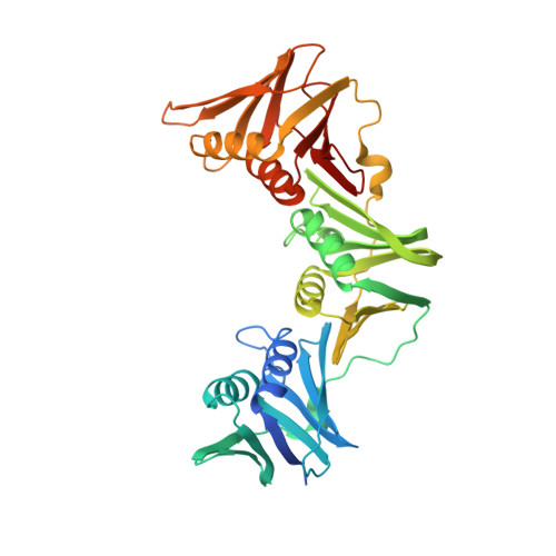
Iterative Structure-Based Optimization of Short Peptides Targeting the Bacterial Sliding Clamp.

Monsarrat, C.Compain, G.Andre, C.Engilberge, S.Martiel, I.Olieric, V.Wolff, P.Brillet, K.Landolfo, M.Silva da Veiga, C.Wagner, J.Guichard, G.Burnouf, D.Y.

(2021) J Med Chem 64: 17063-17078

  • DOI: https://doi.org/10.1021/acs.jmedchem.1c00918
  • Primary Citation of Related Structures:  
    7AZ5, 7AZ6, 7AZ7, 7AZ8, 7AZC, 7AZD, 7AZE, 7AZF, 7AZG, 7AZK, 7AZL

  • PubMed Abstract: 

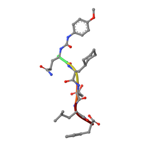
    The bacterial DNA sliding clamp (SC), or replication processivity factor, is a promising target for the development of novel antibiotics. We report a structure-activity relationship study of a new series of peptides interacting within the Escherichia coli SC ( Ec SC) binding pocket. Various modifications were explored including N-alkylation of the peptide bonds, extension of the N-terminal moiety, and introduction of hydrophobic and constrained residues at the C-terminus. In each category, single modifications were identified that increased affinity to Ec SC. A combination of such modifications yielded in several cases to a substantially increased affinity compared to the parent peptides with K d in the range of 30-80 nM. X-ray structure analysis of 11 peptide/ Ec SC co-crystals revealed new interactions at the peptide-protein interface (i.e., stacking interactions, hydrogen bonds, and hydrophobic contacts) that can account for the improved binding. Several compounds among the best binders were also found to be more effective in inhibiting SC-dependent DNA synthesis.


  • Organizational Affiliation

    Université de Bordeaux, CNRS, Bordeaux INP, CBMN, UMR 5248, Institut Européen de Chimie et Biologie, 2 rue Robert Escarpit, F-33607 Pessac, France.


Macromolecules
Find similar proteins by:  (by identity cutoff)  |  3D Structure
Entity ID: 1
MoleculeChains Sequence LengthOrganismDetailsImage
Beta sliding clamp
A, B, C, D
386Escherichia coli 2-427-07_S4_C3Mutation(s): 0 
Gene Names: dnaNAD31_4438
UniProt
Find proteins for P0A988 (Escherichia coli (strain K12))
Explore P0A988 
Go to UniProtKB:  P0A988
Entity Groups  
Sequence Clusters30% Identity50% Identity70% Identity90% Identity95% Identity100% Identity
UniProt GroupP0A988
Sequence Annotations
Expand
  • Reference Sequence

Find similar proteins by:  Sequence   |   3D Structure  

Entity ID: 2
MoleculeChains Sequence LengthOrganismDetailsImage
Peptide 22E [auth H],
F [auth I],
G [auth J],
H [auth K]
6synthetic constructMutation(s): 0 
Sequence Annotations
Expand
  • Reference Sequence
Small Molecules
Modified Residues  1 Unique
IDChains TypeFormula2D DiagramParent
ALC
Query on ALC
E [auth H],
F [auth I],
G [auth J],
H [auth K]
L-PEPTIDE LINKINGC9 H17 N O2ALA
Experimental Data & Validation

Experimental Data

  • Method: X-RAY DIFFRACTION
  • Resolution: 1.77 Å
  • R-Value Free: 0.220 
  • R-Value Work: 0.173 
  • R-Value Observed: 0.175 
  • Space Group: P 1
Unit Cell:
Length ( Å )Angle ( ˚ )
a = 73.809α = 70.9
b = 76.785β = 68.22
c = 79.806γ = 89.14
Software Package:
Software NamePurpose
XDSdata reduction
Aimlessdata scaling
PHASERphasing
BUSTERrefinement
PDB_EXTRACTdata extraction

Structure Validation

View Full Validation Report


Entry History 

Revision History  (Full details and data files)

  • Version 1.0: 2021-12-01
    Type: Initial release
  • Version 1.1: 2022-06-15
    Changes: Database references
  • Version 2.0: 2023-06-21
    Changes: Advisory, Atomic model, Author supporting evidence, Data collection, Database references, Derived calculations, Non-polymer description, Polymer sequence, Structure summary
  • Version 2.1: 2024-02-07
    Changes: Data collection, Derived calculations, Refinement description