7AWN

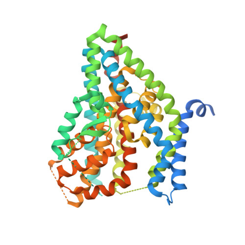
Structure of the thermostabilized EAAT1 cryst mutant in complex with rubidium and barium and the allosteric inhibitor UCPH101


Experimental Data Snapshot

  • Method: X-RAY DIFFRACTION
  • Resolution: 3.92 Å
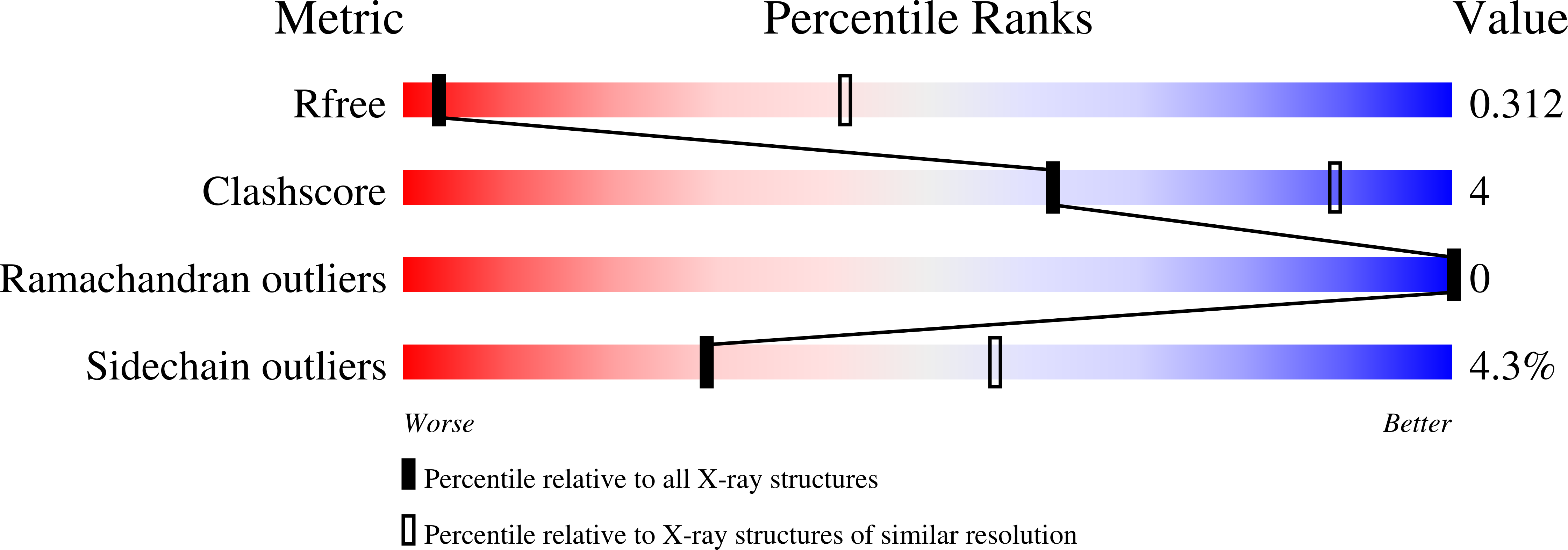
  • R-Value Free: 0.279 
  • R-Value Work: 0.230 
  • R-Value Observed: 0.233 

wwPDB Validation   3D Report Full Report


This is version 1.4 of the entry. See complete history


Literature

The ion-coupling mechanism of human excitatory amino acid transporters.

Canul-Tec, J.C.Kumar, A.Dhenin, J.Assal, R.Legrand, P.Rey, M.Chamot-Rooke, J.Reyes, N.

(2022) EMBO J 41: e108341-e108341

  • DOI: https://doi.org/10.15252/embj.2021108341
  • Primary Citation of Related Structures:  
    7AWL, 7AWM, 7AWN, 7AWP, 7AWQ, 7NPW

  • PubMed Abstract: 

    Excitatory amino acid transporters (EAATs) maintain glutamate gradients in the brain essential for neurotransmission and to prevent neuronal death. They use ionic gradients as energy source and co-transport transmitter into the cytoplasm with Na + and H + , while counter-transporting K + to re-initiate the transport cycle. However, the molecular mechanisms underlying ion-coupled transport remain incompletely understood. Here, we present 3D X-ray crystallographic and cryo-EM structures, as well as thermodynamic analysis of human EAAT1 in different ion bound conformations, including elusive counter-transport ion bound states. Binding energies of Na + and H + , and unexpectedly Ca 2+ , are coupled to neurotransmitter binding. Ca 2+ competes for a conserved Na + site, suggesting a regulatory role for Ca 2+ in glutamate transport at the synapse, while H + binds to a conserved glutamate residue stabilizing substrate occlusion. The counter-transported ion binding site overlaps with that of glutamate, revealing the K + -based mechanism to exclude the transmitter during the transport cycle and to prevent its neurotoxic release on the extracellular side.


  • Organizational Affiliation

    Membrane Protein Mechanisms Unit, Institut Pasteur, Paris, France.


Macromolecules
Find similar proteins by:  (by identity cutoff)  |  3D Structure
Entity ID: 1
MoleculeChains Sequence LengthOrganismDetailsImage
Excitatory amino acid transporter 1,Neutral amino acid transporter B(0),Excitatory amino acid transporter 1522Homo sapiensMutation(s): 73 
Gene Names: SLC1A3EAAT1GLASTGLAST1SLC1A5ASCT2M7V1RDRRDRC
Membrane Entity: Yes 
UniProt & NIH Common Fund Data Resources
Find proteins for Q15758 (Homo sapiens)
Explore Q15758 
Go to UniProtKB:  Q15758
PHAROS:  Q15758
GTEx:  ENSG00000105281 
Find proteins for P43003 (Homo sapiens)
Explore P43003 
Go to UniProtKB:  P43003
PHAROS:  P43003
GTEx:  ENSG00000079215 
Entity Groups  
Sequence Clusters30% Identity50% Identity70% Identity90% Identity95% Identity100% Identity
UniProt GroupsP43003Q15758
Sequence Annotations
Expand
  • Reference Sequence
Experimental Data & Validation

Experimental Data

  • Method: X-RAY DIFFRACTION
  • Resolution: 3.92 Å
  • R-Value Free: 0.279 
  • R-Value Work: 0.230 
  • R-Value Observed: 0.233 
  • Space Group: P 63
Unit Cell:
Length ( Å )Angle ( ˚ )
a = 124.236α = 90
b = 124.236β = 90
c = 91.22γ = 120
Software Package:
Software NamePurpose
XDSdata reduction
Aimlessdata scaling
PHASERphasing
BUSTERrefinement
PDB_EXTRACTdata extraction

Structure Validation

View Full Validation Report


Entry History & Funding Information

Deposition Data


Funding OrganizationLocationGrant Number
European Research Council (ERC)France309657

Revision History  (Full details and data files)

  • Version 1.0: 2021-10-13
    Type: Initial release
  • Version 1.1: 2021-12-01
    Changes: Database references
  • Version 1.2: 2022-01-12
    Changes: Database references
  • Version 1.3: 2022-01-19
    Changes: Database references
  • Version 1.4: 2024-01-31
    Changes: Data collection, Refinement description