7ATM

Structure of P. aeruginosa PBP3 in complex with a phenyl boronic acid (Compound 1)


Experimental Data Snapshot

  • Method: X-RAY DIFFRACTION
  • Resolution: 1.58 Å
  • R-Value Free: 0.225 
  • R-Value Work: 0.191 
  • R-Value Observed: 0.192 

wwPDB Validation   3D Report Full Report


Ligand Structure Quality Assessment 


This is version 1.2 of the entry. See complete history


Literature

High-Throughput Crystallography Reveals Boron-Containing Inhibitors of a Penicillin-Binding Protein with Di- and Tricovalent Binding Modes.

Newman, H.Krajnc, A.Bellini, D.Eyermann, C.J.Boyle, G.A.Paterson, N.G.McAuley, K.E.Lesniak, R.Gangar, M.von Delft, F.Brem, J.Chibale, K.Schofield, C.J.Dowson, C.G.

(2021) J Med Chem 64: 11379-11394

  • DOI: https://doi.org/10.1021/acs.jmedchem.1c00717
  • Primary Citation of Related Structures:  
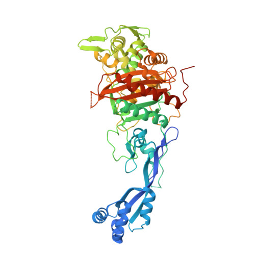
    7ATM, 7ATO, 7ATW, 7ATX, 7AU0, 7AU1, 7AU8, 7AU9, 7AUB, 7AUH

  • PubMed Abstract: 

    The effectiveness of β-lactam antibiotics is increasingly compromised by β-lactamases. Boron-containing inhibitors are potent serine-β-lactamase inhibitors, but the interactions of boron-based compounds with the penicillin-binding protein (PBP) β-lactam targets have not been extensively studied. We used high-throughput X-ray crystallography to explore reactions of a boron-containing fragment set with the Pseudomonas aeruginosa PBP3 (PaPBP3). Multiple crystal structures reveal that boronic acids react with PBPs to give tricovalently linked complexes bonded to Ser294, Ser349, and Lys484 of PaPBP3; benzoxaboroles react with PaPBP3 via reaction with two nucleophilic serines (Ser294 and Ser349) to give dicovalently linked complexes; and vaborbactam reacts to give a monocovalently linked complex. Modifications of the benzoxaborole scaffold resulted in a moderately potent inhibition of PaPBP3, though no antibacterial activity was observed. Overall, the results further evidence the potential for the development of new classes of boron-based antibiotics, which are not compromised by β-lactamase-driven resistance.


  • Organizational Affiliation

    School of Life Sciences, University of Warwick, Coventry CV4 7AL, U.K.


Macromolecules
Find similar proteins by:  (by identity cutoff)  |  3D Structure
Entity ID: 1
MoleculeChains Sequence LengthOrganismDetailsImage
Peptidoglycan D,D-transpeptidase FtsI537Pseudomonas aeruginosa PAO1Mutation(s): 0 
Gene Names: ftsIpbpBPA4418
EC: 3.4.16.4
UniProt
Find proteins for G3XD46 (Pseudomonas aeruginosa (strain ATCC 15692 / DSM 22644 / CIP 104116 / JCM 14847 / LMG 12228 / 1C / PRS 101 / PAO1))
Explore G3XD46 
Go to UniProtKB:  G3XD46
Entity Groups  
Sequence Clusters30% Identity50% Identity70% Identity90% Identity95% Identity100% Identity
UniProt GroupG3XD46
Sequence Annotations
Expand
  • Reference Sequence
Experimental Data & Validation

Experimental Data

  • Method: X-RAY DIFFRACTION
  • Resolution: 1.58 Å
  • R-Value Free: 0.225 
  • R-Value Work: 0.191 
  • R-Value Observed: 0.192 
  • Space Group: P 21 21 21
Unit Cell:
Length ( Å )Angle ( ˚ )
a = 69.845α = 90
b = 80.951β = 90
c = 88.8γ = 90
Software Package:
Software NamePurpose
XDSdata reduction
Aimlessdata scaling
PHASERphasing
REFMACrefinement
PDB_EXTRACTdata extraction
STARANISOdata processing

Structure Validation

View Full Validation Report


Ligand Structure Quality Assessment 


Entry History & Funding Information

Deposition Data


Funding OrganizationLocationGrant Number
Medical Research Council (MRC, United Kingdom)United Kingdom--

Revision History  (Full details and data files)

  • Version 1.0: 2021-08-11
    Type: Initial release
  • Version 1.1: 2021-09-29
    Changes: Data collection, Database references
  • Version 1.2: 2024-01-31
    Changes: Data collection, Refinement description