


Crystal Structure of an Anti-Plasminogen Activator Inhibitor-1 (PAI-1) scFv antibody fragment (scFv-33H1F7)
Sillen, M., Weeks, S.D., Strelkov, S.V., Declerck, P.J.To be published.
Experimental Data Snapshot
Starting Model: experimental
View more details
wwPDB Validation 3D Report Full Report

Entity ID: 1 | |||||
|---|---|---|---|---|---|
| Molecule | Chains | Sequence Length | Organism | Details | Image |
| scFv-33H1F7 | A [auth B], B [auth A], C, D | 242 | Mus musculus | Mutation(s): 0 | 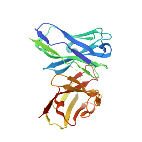 |
| Ligands 1 Unique | |||||
|---|---|---|---|---|---|
| ID | Chains | Name / Formula / InChI Key | 2D Diagram | 3D Interactions | |
| SO4 Download:Ideal Coordinates CCD File | E [auth B] F [auth B] G [auth A] H [auth A] I [auth C] | SULFATE ION O4 S QAOWNCQODCNURD-UHFFFAOYSA-L |  | ||
| Length ( Å ) | Angle ( ˚ ) |
|---|---|
| a = 190.398 | α = 90 |
| b = 55.577 | β = 90.28 |
| c = 102.493 | γ = 90 |
| Software Name | Purpose |
|---|---|
| PHENIX | refinement |
| Aimless | data scaling |
| PDB_EXTRACT | data extraction |
| XDS | data reduction |
| PHASER | phasing |
| Funding Organization | Location | Grant Number |
|---|---|---|
| Research Foundation - Flanders (FWO) | Belgium | 11ZQ918N |
| Research Foundation - Flanders (FWO) | Belgium | G072915N |


