Picomolar FKBP inhibitors enabled by a single water-displacing methyl group in bicyclic [4.3.1] aza-amides.
Kolos, J.M., Pomplun, S., Jung, S., Riess, B., Purder, P.L., Voll, A.M., Merz, S., Gnatzy, M., Geiger, T.M., Quist-Lokken, I., Jatzlau, J., Knaus, P., Holien, T., Bracher, A., Meyners, C., Czodrowski, P., Krewald, V., Hausch, F.(2021) Chem Sci 12: 14758-14765
- PubMed: 34820091 Search on PubMedSearch on PubMed Central
- DOI: https://doi.org/10.1039/d1sc04638a
- Primary Citation Related Structures:
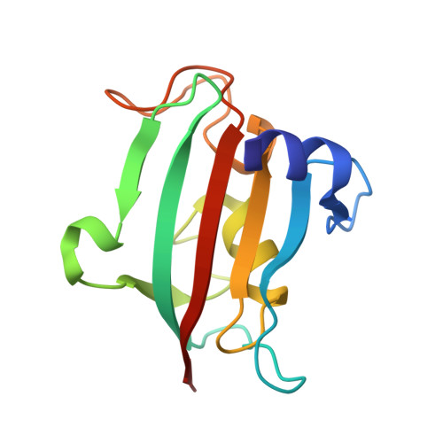
7APQ, 7APS, 7APT, 7APW - PubMed Abstract:
Methyl groups can have profound effects in drug discovery but the underlying mechanisms are diverse and incompletely understood. Here we report the stereospecific effect of a single, solvent-exposed methyl group in bicyclic [4.3.1] aza-amides, robustly leading to a 2 to 10-fold increase in binding affinity for FK506-binding proteins (FKBPs). This resulted in the most potent and efficient FKBP ligands known to date. By a combination of co-crystal structures, isothermal titration calorimetry (ITC), density-functional theory (DFT), and 3D reference interaction site model (3D-RISM) calculations we elucidated the origin of the observed affinity boost, which was purely entropically driven and relied on the displacement of a water molecule at the protein-ligand-bulk solvent interface. The best compounds potently occupied FKBPs in cells and enhanced bone morphogenic protein (BMP) signaling. Our results show how subtle manipulation of the solvent network can be used to design atom-efficient ligands for difficult, solvent-exposed binding pockets.
- Department of Chemistry, Technical University of Darmstadt Alarich-Weiss-Straße 4 64293 Darmstadt Germany felix.hausch@tu-darmstadt.de.
Organizational Affiliation: