7ALF | pdb_00007alf

The dimethylated RSL - sulfonato-calix[8]arene complex, P3 form, acetate pH 4.0


Experimental Data Snapshot

  • Method: X-RAY DIFFRACTION
  • Resolution: 1.26 Å
  • R-Value Free: 
    0.162 (Depositor), 0.162 (DCC) 
  • R-Value Work: 
    0.145 (Depositor), 0.145 (DCC) 
  • R-Value Observed: 
    0.145 (Depositor) 

Starting Model: experimental
View more details

wwPDB Validation 3D Report Full Report

Validation slider image for 7ALF

Ligand Structure Quality Assessment 


This is version 1.1 of the entry. See complete history

Literature

Facile Fabrication of Protein-Macrocycle Frameworks.

Ramberg, K.O.Engilberge, S.Skorek, T.Crowley, P.B.

(2021) J Am Chem Soc 143: 1896-1907

  • DOI: https://doi.org/10.1021/jacs.0c10697
  • Primary Citation Related Structures: 
    6Z5G, 6Z5M, 6Z5P, 6Z5Q, 6Z5W, 6Z5X, 6Z5Z, 6Z60, 6Z62, 7ALF, 7ALG

  • PubMed Abstract: 

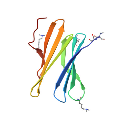
    Precisely defined protein aggregates, as exemplified by crystals, have applications in functional materials. Consequently, engineered protein assembly is a rapidly growing field. Anionic calix[n]arenes are useful scaffolds that can mold to cationic proteins and induce oligomerization and assembly. Here, we describe protein-calixarene composites obtained via cocrystallization of commercially available sulfonato-calix[8]arene ( sclx 8 ) with the symmetric and "neutral" protein RSL. Cocrystallization occurred across a wide range of conditions and protein charge states, from pH 2.2-9.5, resulting in three crystal forms. Cationization of the protein surface at pH ∼ 4 drives calixarene complexation and yielded two types of porous frameworks with pore diameters >3 nm. Both types of framework provide evidence of protein encapsulation by the calixarene. Calixarene-masked proteins act as nodes within the frameworks, displaying octahedral-type coordination in one case. The other framework formed millimeter-scale crystals within hours, without the need for precipitants or specialized equipment. NMR experiments revealed macrocycle - modulated side chain p K a values and suggested a mechanism for pH-triggered assembly. The same low pH framework was generated at high pH with a permanently cationic arginine-enriched RSL variant. Finally, in addition to protein framework fabrication, sclx 8 enables de novo structure determination.


  • Organizational Affiliation
    • School of Chemistry, National University of Ireland Galway, University Road, Galway, H91 TK33, Ireland.

Macromolecule Content 

  • Total Structure Weight: 23.57 kDa 
  • Atom Count: 2,011 
  • Modeled Residue Count: 180 
  • Deposited Residue Count: 180 
  • Unique protein chains: 1

Macromolecules

Find similar proteins by:|  3D Structure
Entity ID: 1
MoleculeChains  Sequence LengthOrganismDetailsImage
Fucose-binding lectin protein
A, B
90Ralstonia solanacearumMutation(s): 0 
Gene Names: RSP795_21825RSP799_05830RUN39_v1_50103
UniProt
Find proteins for A0A0S4TLR1 (Ralstonia solanacearum)
Explore A0A0S4TLR1 
Go to UniProtKB:  A0A0S4TLR1
Entity Groups
Sequence Clusters30% Identity50% Identity70% Identity90% Identity95% Identity100% Identity
UniProt GroupA0A0S4TLR1
Sequence Annotations
Expand
Reference Sequence

Small Molecules

Ligands 3 Unique
IDChains Name / Formula / InChI Key2D Diagram3D Interactions
EVB
(Subject of Investigation/LOI)

Query on EVB


Download:Ideal Coordinates CCD File
C [auth A],
G [auth B]
sulfonato-calix[8]arene
C56 H48 O32 S8
KCEGJGDGMRAJEP-UHFFFAOYSA-N
BDF

Query on BDF


Download:Ideal Coordinates CCD File
E [auth A],
F [auth A],
I [auth B],
J [auth B]
beta-D-fructopyranose
C6 H12 O6
LKDRXBCSQODPBY-ARQDHWQXSA-N
GOL

Query on GOL


Download:Ideal Coordinates CCD File
D [auth A],
H [auth B]
GLYCEROL
C3 H8 O3
PEDCQBHIVMGVHV-UHFFFAOYSA-N
Modified Residues  1 Unique
IDChains TypeFormula2D DiagramParent
MLY
Query on MLY
A, B
L-PEPTIDE LINKINGC8 H18 N2 O2LYS

Experimental Data & Validation

Experimental Data

  • Method: X-RAY DIFFRACTION
  • Resolution: 1.26 Å
  • R-Value Free:  0.162 (Depositor), 0.162 (DCC) 
  • R-Value Work:  0.145 (Depositor), 0.145 (DCC) 
  • R-Value Observed: 0.145 (Depositor) 
Space Group: P 3
Unit Cell:
Length ( Å )Angle ( ˚ )
a = 59.478α = 90
b = 59.478β = 90
c = 64.72γ = 120
Software Package:
Software NamePurpose
PHENIXrefinement
PDB_EXTRACTdata extraction
XDSdata reduction
Aimlessdata scaling
PHASERphasing

Structure Validation

View Full Validation Report


Ligand Structure Quality Assessment 


Entry History 

& Funding Information

Deposition Data


Funding OrganizationLocationGrant Number
Science Foundation IrelandIreland13/CDA/2168

Revision History  (Full details and data files)

  • Version 1.0: 2021-04-07
    Type: Initial release
  • Version 1.1: 2024-01-31
    Changes: Advisory, Data collection, Database references, Refinement description