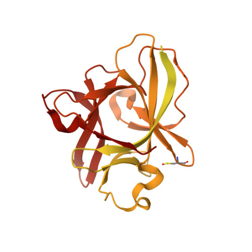
7AL7

The Crystal Structure of Human IL-18 in Complex With Human IL-18 Binding Protein


Experimental Data Snapshot

  • Method: X-RAY DIFFRACTION
  • Resolution: 1.80 Å
  • R-Value Free: 0.232 
  • R-Value Work: 0.209 
  • R-Value Observed: 0.211 

wwPDB Validation   3D Report Full Report


Ligand Structure Quality Assessment 


This is version 1.3 of the entry. See complete history


Literature

Structural basis of human IL-18 sequestration by the decoy receptor IL-18 binding protein in inflammation and tumor immunity.

Detry, S.Andries, J.Bloch, Y.Gabay, C.Clancy, D.M.Savvides, S.N.

(2022) J Biol Chem 298: 101908-101908

  • DOI: https://doi.org/10.1016/j.jbc.2022.101908
  • Primary Citation of Related Structures:  
    7AL7

  • PubMed Abstract: 

    Human Interleukin-18 (IL-18) is an omnipresent proinflammatory cytokine of the IL-1 family with central roles in autoimmune and inflammatory diseases and serves as a staple biomarker in the evaluation of inflammation in physiology and disease, including the inflammatory phase of COVID-19. The sequestration of IL-18 by its soluble decoy receptor IL-18-Binding Protein (IL-18BP) is critical to the regulation of IL-18 activity. Since an imbalance in expression and circulating levels of IL-18 is associated with disease, structural insights into how IL-18BP outcompetes binding of IL-18 by its cognate cell-surface receptors are highly desirable; however, the structure of human IL-18BP in complex with IL-18 has been elusive. Here, we elucidate the sequestration mechanism of human IL-18 mediated by IL-18BP based on the crystal structure of the IL-18:IL-18BP complex. These detailed structural snapshots reveal the interaction landscape leading to the ultra-high affinity of IL-18BP toward IL-18 and identify substantial differences with respect to previously characterized complexes of IL-18 with IL-18BP of viral origin. Furthermore, our structure captured a fortuitous higher-order assembly between IL-18 and IL-18BP coordinated by a disulfide-bond distal to the binding surface connecting IL-18 and IL-18BP molecules from different complexes, resulting in a novel tetramer with 2:2 stoichiometry. This tetrapartite assembly was found to restrain IL-18 activity more effectively than the canonical 1:1 complex. Collectively, our findings provide a framework for innovative, structure-driven therapeutic strategies and further functional interrogation of IL-18 in physiology and disease.


  • Organizational Affiliation

    Unit for Structural Biology, Department of Biochemistry and Microbiology, Ghent University, Ghent, Belgium; Unit for Structural Biology, VIB Center for Inflammation Research, Ghent, Belgium.


Macromolecules
Find similar proteins by:  (by identity cutoff)  |  3D Structure
Entity ID: 1
MoleculeChains Sequence LengthOrganismDetailsImage
Interleukin-18-binding protein172Homo sapiensMutation(s): 0 
Gene Names: IL18BP
UniProt & NIH Common Fund Data Resources
Find proteins for O95998 (Homo sapiens)
Explore O95998 
Go to UniProtKB:  O95998
PHAROS:  O95998
GTEx:  ENSG00000137496 
Entity Groups  
Sequence Clusters30% Identity50% Identity70% Identity90% Identity95% Identity100% Identity
UniProt GroupO95998
Sequence Annotations
Expand
  • Reference Sequence
Find similar proteins by:  (by identity cutoff)  |  3D Structure
Entity ID: 2
MoleculeChains Sequence LengthOrganismDetailsImage
Glutathione S-transferase class-mu 26 kDa isozyme,Interleukin-18390Schistosoma japonicumHomo sapiens
This entity is chimeric
Mutation(s): 0 
EC: 2.5.1.18
UniProt & NIH Common Fund Data Resources
Find proteins for Q14116 (Homo sapiens)
Explore Q14116 
Go to UniProtKB:  Q14116
PHAROS:  Q14116
GTEx:  ENSG00000150782 
Find proteins for P08515 (Schistosoma japonicum)
Explore P08515 
Go to UniProtKB:  P08515
Entity Groups  
Sequence Clusters30% Identity50% Identity70% Identity90% Identity95% Identity100% Identity
UniProt GroupsQ14116P08515
Sequence Annotations
Expand
  • Reference Sequence
Small Molecules
Modified Residues  2 Unique
IDChains TypeFormula2D DiagramParent
PCA
Query on PCA
A
L-PEPTIDE LINKINGC5 H7 N O3GLN
CSO
Query on CSO
B
L-PEPTIDE LINKINGC3 H7 N O3 SCYS
Experimental Data & Validation

Experimental Data

  • Method: X-RAY DIFFRACTION
  • Resolution: 1.80 Å
  • R-Value Free: 0.232 
  • R-Value Work: 0.209 
  • R-Value Observed: 0.211 
  • Space Group: C 1 2 1
Unit Cell:
Length ( Å )Angle ( ˚ )
a = 109.815α = 90
b = 44.52β = 99.86
c = 60.276γ = 90
Software Package:
Software NamePurpose
PHENIXrefinement
PDB_EXTRACTdata extraction
XDSdata reduction
XSCALEdata scaling
PHASERphasing
STARANISOdata scaling

Structure Validation

View Full Validation Report


Ligand Structure Quality Assessment 


Entry History & Funding Information

Deposition Data


Funding OrganizationLocationGrant Number
European Union (EU)European Union779295

Revision History  (Full details and data files)

  • Version 1.0: 2022-04-20
    Type: Initial release
  • Version 1.1: 2022-04-27
    Changes: Database references
  • Version 1.2: 2022-05-25
    Changes: Database references
  • Version 1.3: 2024-01-31
    Changes: Data collection, Refinement description