7AKS

Human ADP-ribosylserine hydrolase ARH3 mutant E41A in complex with H2B-S7-mar peptide


Experimental Data Snapshot

  • Method: X-RAY DIFFRACTION
  • Resolution: 1.86 Å
  • R-Value Free: 0.195 
  • R-Value Work: 0.169 

wwPDB Validation   3D Report Full Report


Ligand Structure Quality Assessment 


This is version 1.2 of the entry. See complete history


Literature

Mechanistic insights into the three steps of poly(ADP-ribosylation) reversal.

Rack, J.G.M.Liu, Q.Zorzini, V.Voorneveld, J.Ariza, A.Honarmand Ebrahimi, K.Reber, J.M.Krassnig, S.C.Ahel, D.van der Marel, G.A.Mangerich, A.McCullagh, J.S.O.Filippov, D.V.Ahel, I.

(2021) Nat Commun 12: 4581-4581

  • DOI: https://doi.org/10.1038/s41467-021-24723-3
  • Primary Citation of Related Structures:  
    7AKR, 7AKS, 7AQM, 7ARW

  • PubMed Abstract: 

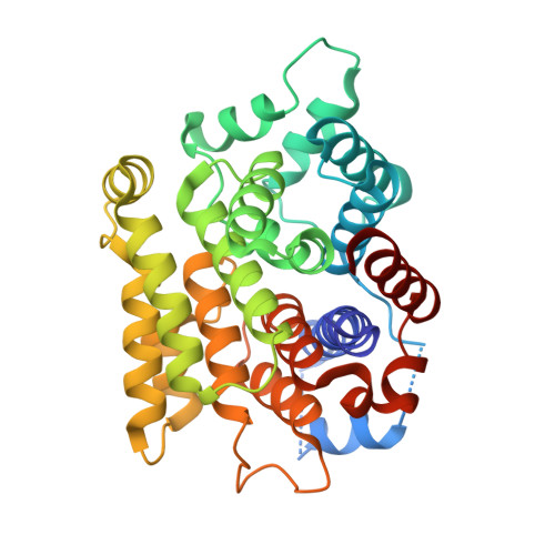
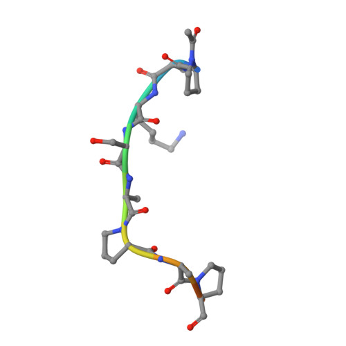
    Poly(ADP-ribosyl)ation (PAR) is a versatile and complex posttranslational modification composed of repeating units of ADP-ribose arranged into linear or branched polymers. This scaffold is linked to the regulation of many of cellular processes including the DNA damage response, alteration of chromatin structure and Wnt signalling. Despite decades of research, the principles and mechanisms underlying all steps of PAR removal remain actively studied. In this work, we synthesise well-defined PAR branch point molecules and demonstrate that PARG, but not ARH3, can resolve this distinct PAR architecture. Structural analysis of ARH3 in complex with dimeric ADP-ribose as well as an ADP-ribosylated peptide reveal the molecular basis for the hydrolysis of linear and terminal ADP-ribose linkages. We find that ARH3-dependent hydrolysis requires both rearrangement of a catalytic glutamate and induction of an unusual, square-pyramidal magnesium coordination geometry.


  • Organizational Affiliation

    Sir William Dunn School of Pathology, University of Oxford, Oxford, UK.


Macromolecules
Find similar proteins by:  (by identity cutoff)  |  3D Structure
Entity ID: 1
MoleculeChains Sequence LengthOrganismDetailsImage
ADP-ribose glycohydrolase ARH3A [auth AAA],
C [auth CCC],
E [auth EEE],
G [auth GGG]
349Homo sapiensMutation(s): 1 
Gene Names: ADPRSADPRHL2ARH3
EC: 3.5.1 (PDB Primary Data), 3.2.1.143 (PDB Primary Data), 3.2.2 (PDB Primary Data)
UniProt & NIH Common Fund Data Resources
Find proteins for Q9NX46 (Homo sapiens)
Explore Q9NX46 
Go to UniProtKB:  Q9NX46
PHAROS:  Q9NX46
GTEx:  ENSG00000116863 
Entity Groups  
Sequence Clusters30% Identity50% Identity70% Identity90% Identity95% Identity100% Identity
UniProt GroupQ9NX46
Sequence Annotations
Expand
  • Reference Sequence

Find similar proteins by:  Sequence   |   3D Structure  

Entity ID: 2
MoleculeChains Sequence LengthOrganismDetailsImage
modified peptideB [auth BaB],
D [auth DaD],
F [auth FaF],
H [auth HaH]
11Homo sapiensMutation(s): 0 
EC: 3.5.1 (PDB Primary Data), 3.2.1.143 (PDB Primary Data), 3.2.2 (PDB Primary Data)
Entity Groups  
Sequence Clusters30% Identity50% Identity70% Identity90% Identity95% Identity100% Identity
Sequence Annotations
Expand
  • Reference Sequence
Small Molecules
Ligands 4 Unique
IDChains Name / Formula / InChI Key2D Diagram3D Interactions
AR6 (Subject of Investigation/LOI)
Query on AR6

Download Ideal Coordinates CCD File 
CB [auth FaF],
LA [auth DaD],
PB [auth HaH],
T [auth BaB]
[(2R,3S,4R,5R)-5-(6-AMINOPURIN-9-YL)-3,4-DIHYDROXY-OXOLAN-2-YL]METHYL [HYDROXY-[[(2R,3S,4R,5S)-3,4,5-TRIHYDROXYOXOLAN-2-YL]METHOXY]PHOSPHORYL] HYDROGEN PHOSPHATE
C15 H23 N5 O14 P2
SRNWOUGRCWSEMX-ZQSHOCFMSA-N
EDO
Query on EDO

Download Ideal Coordinates CCD File 
AA [auth CCC]
BA [auth CCC]
CA [auth CCC]
FB [auth GGG]
GB [auth GGG]
AA [auth CCC],
BA [auth CCC],
CA [auth CCC],
FB [auth GGG],
GB [auth GGG],
HB [auth GGG],
K [auth AAA],
L [auth AAA],
M [auth AAA],
OA [auth EEE],
PA [auth EEE],
QA [auth EEE],
RA [auth EEE],
SA [auth EEE],
TA [auth EEE],
UA [auth EEE],
X [auth CCC],
Y [auth CCC],
Z [auth CCC]
1,2-ETHANEDIOL
C2 H6 O2
LYCAIKOWRPUZTN-UHFFFAOYSA-N
ACT
Query on ACT

Download Ideal Coordinates CCD File 
AB [auth EEE]
BB [auth EEE]
DA [auth CCC]
EA [auth CCC]
FA [auth CCC]
AB [auth EEE],
BB [auth EEE],
DA [auth CCC],
EA [auth CCC],
FA [auth CCC],
GA [auth CCC],
HA [auth CCC],
IA [auth CCC],
IB [auth GGG],
JA [auth CCC],
JB [auth GGG],
KA [auth CCC],
KB [auth GGG],
LB [auth GGG],
MB [auth GGG],
N [auth AAA],
NB [auth GGG],
O [auth AAA],
OB [auth GGG],
P [auth AAA],
Q [auth AAA],
R [auth AAA],
S [auth AAA],
VA [auth EEE],
WA [auth EEE],
XA [auth EEE],
YA [auth EEE],
ZA [auth EEE]
ACETATE ION
C2 H3 O2
QTBSBXVTEAMEQO-UHFFFAOYSA-M
MG
Query on MG

Download Ideal Coordinates CCD File 
DB [auth GGG]
EB [auth GGG]
I [auth AAA]
J [auth AAA]
MA [auth EEE]
DB [auth GGG],
EB [auth GGG],
I [auth AAA],
J [auth AAA],
MA [auth EEE],
NA [auth EEE],
U [auth CCC],
V [auth CCC],
W [auth CCC]
MAGNESIUM ION
Mg
JLVVSXFLKOJNIY-UHFFFAOYSA-N
Modified Residues  1 Unique
IDChains TypeFormula2D DiagramParent
N7P
Query on N7P
B [auth BaB],
D [auth DaD],
F [auth FaF],
H [auth HaH]
L-PEPTIDE LINKINGC7 H11 N O3PRO
Experimental Data & Validation

Experimental Data

  • Method: X-RAY DIFFRACTION
  • Resolution: 1.86 Å
  • R-Value Free: 0.195 
  • R-Value Work: 0.169 
  • Space Group: P 21 21 2
Unit Cell:
Length ( Å )Angle ( ˚ )
a = 123.36α = 90
b = 158.638β = 90
c = 74.92γ = 90
Software Package:
Software NamePurpose
REFMACrefinement
DIALSdata reduction
Aimlessdata scaling
PHENIXphasing

Structure Validation

View Full Validation Report


Ligand Structure Quality Assessment 


Entry History & Funding Information

Deposition Data

  • Released Date: 2021-06-16 
  • Deposition Author(s): Ariza, A.

Funding OrganizationLocationGrant Number
Wellcome TrustUnited Kingdom101794
Wellcome TrustUnited Kingdom210634

Revision History  (Full details and data files)

  • Version 1.0: 2021-06-16
    Type: Initial release
  • Version 1.1: 2021-10-06
    Changes: Data collection, Database references
  • Version 1.2: 2024-01-31
    Changes: Data collection, Derived calculations, Refinement description, Source and taxonomy