7AHS | pdb_00007ahs

titin-N2A Ig81-Ig83


Experimental Data Snapshot

  • Method: X-RAY DIFFRACTION
  • Resolution: 2.05 Å
  • R-Value Free: 
    0.235 (Depositor), 0.236 (DCC) 
  • R-Value Work: 
    0.183 (Depositor), 0.184 (DCC) 
  • R-Value Observed: 
    0.185 (Depositor) 

Starting Model: experimental
View more details

wwPDB Validation 3D Report Full Report

Validation slider image for 7AHS

This is version 1.2 of the entry. See complete history

Literature

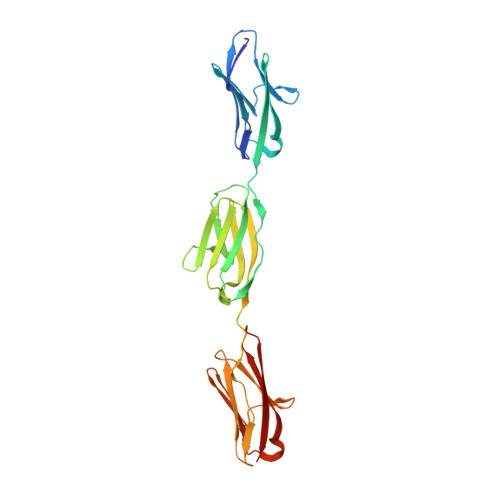
The N2A region of titin has a unique structural configuration.

Stronczek, C.Lange, S.Bullard, B.Wolniak, S.Borgeson, E.Mayans, O.Fleming, J.R.

(2021) J Gen Physiol 153

  • DOI: https://doi.org/10.1085/jgp.202012766
  • Primary Citation Related Structures: 
    7AHS

  • PubMed Abstract: 

    The N2A segment of titin is a main signaling hub in the sarcomeric I-band that recruits various signaling factors and processing enzymes. It has also been proposed to play a role in force production through its Ca2+-regulated association with actin. However, the molecular basis by which N2A performs these functions selectively within the repetitive and extensive titin chain remains poorly understood. Here, we analyze the structure of N2A components and their association with F-actin. Specifically, we characterized the structure of its Ig domains by elucidating the atomic structure of the I81-I83 tandem using x-ray crystallography and computing a homology model for I80. Structural data revealed these domains to present heterogeneous and divergent Ig folds, where I81 and I83 have unique loop structures. Notably, the I81-I83 tandem has a distinct rotational chain arrangement that confers it a unique multi-domain topography. However, we could not identify specific Ca2+-binding sites in these Ig domains, nor evidence of the association of titin N2A components with F-actin in transfected C2C12 myoblasts or C2C12-derived myotubes. In addition, F-actin cosedimentation assays failed to reveal binding to N2A. We conclude that N2A has a unique architecture that predictably supports its selective recruitment of binding partners in signaling, but that its mechanical role through interaction with F-actin awaits validation.


  • Organizational Affiliation
    • Department of Biology, University of Konstanz, Konstanz, Germany.

Macromolecule Content 

  • Total Structure Weight: 128.1 kDa 
  • Atom Count: 9,852 
  • Modeled Residue Count: 1,086 
  • Deposited Residue Count: 1,092 
  • Unique protein chains: 1

Macromolecules

Find similar proteins by:|  3D Structure
Entity ID: 1
MoleculeChains  Sequence LengthOrganismDetailsImage
Isoform 5 of Titin
A, B, C, D
273Homo sapiensMutation(s): 0 
Gene Names: TTN
EC: 2.7.11.1
UniProt & NIH Common Fund Data Resources
Find proteins for Q8WZ42 (Homo sapiens)
Explore Q8WZ42 
Go to UniProtKB:  Q8WZ42
PHAROS:  Q8WZ42
GTEx:  ENSG00000155657 
Entity Groups
Sequence Clusters30% Identity50% Identity70% Identity90% Identity95% Identity100% Identity
UniProt GroupQ8WZ42
Sequence Annotations
Expand
Reference Sequence

Small Molecules

Ligands 3 Unique
IDChains Name / Formula / InChI Key2D Diagram3D Interactions
SO4

Query on SO4


Download:Ideal Coordinates CCD File
DA [auth D]
EA [auth D]
L [auth B]
M [auth B]
N [auth B]
DA [auth D],
EA [auth D],
L [auth B],
M [auth B],
N [auth B],
O [auth B],
P [auth B],
Q [auth B],
Y [auth C],
Z [auth C]
SULFATE ION
O4 S
QAOWNCQODCNURD-UHFFFAOYSA-L
GOL

Query on GOL


Download:Ideal Coordinates CCD File
BA [auth C]GLYCEROL
C3 H8 O3
PEDCQBHIVMGVHV-UHFFFAOYSA-N
EDO

Query on EDO


Download:Ideal Coordinates CCD File
AA [auth C]
CA [auth D]
E [auth A]
F [auth A]
FA [auth D]
AA [auth C],
CA [auth D],
E [auth A],
F [auth A],
FA [auth D],
G [auth A],
GA [auth D],
H [auth A],
HA [auth D],
I [auth A],
J [auth B],
K [auth B],
R [auth B],
S [auth B],
T [auth B],
U [auth B],
V [auth B],
W [auth C],
X [auth C]
1,2-ETHANEDIOL
C2 H6 O2
LYCAIKOWRPUZTN-UHFFFAOYSA-N

Experimental Data & Validation

Experimental Data

  • Method: X-RAY DIFFRACTION
  • Resolution: 2.05 Å
  • R-Value Free:  0.235 (Depositor), 0.236 (DCC) 
  • R-Value Work:  0.183 (Depositor), 0.184 (DCC) 
  • R-Value Observed: 0.185 (Depositor) 
Space Group: P 21 21 21
Unit Cell:
Length ( Å )Angle ( ˚ )
a = 85.203α = 90
b = 87.336β = 90
c = 161.583γ = 90
Software Package:
Software NamePurpose
PHENIXrefinement
XSCALEdata reduction
XDSdata scaling
PHASERphasing
ARP/wARPmodel building

Structure Validation

View Full Validation Report


Entry History 

Deposition Data

Revision History  (Full details and data files)

  • Version 1.0: 2021-03-24
    Type: Initial release
  • Version 1.1: 2021-04-21
    Changes: Advisory, Database references, Derived calculations
  • Version 1.2: 2024-01-31
    Changes: Advisory, Data collection, Database references, Refinement description