7AHQ

Cryo-EM structure of F-actin stabilized by trans-optoJASP-8


Experimental Data Snapshot

  • Method: ELECTRON MICROSCOPY
  • Resolution: 3.60 Å
  • Aggregation State: FILAMENT 
  • Reconstruction Method: SINGLE PARTICLE 

wwPDB Validation   3D Report Full Report


This is version 1.1 of the entry. See complete history


Literature

Cryo-EM Resolves Molecular Recognition Of An Optojasp Photoswitch Bound To Actin Filaments In Both Switch States.

Pospich, S.Kullmer, F.Nasufovic, V.Funk, J.Belyy, A.Bieling, P.Arndt, H.D.Raunser, S.

(2021) Angew Chem Int Ed Engl 60: 8678-8682

  • DOI: https://doi.org/10.1002/anie.202013193
  • Primary Citation of Related Structures:  
    7AHN, 7AHQ

  • PubMed Abstract: 

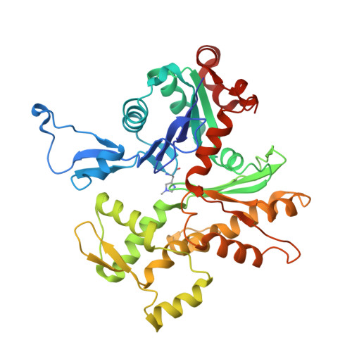
    Actin is essential for key processes in all eukaryotic cells. Cellpermeable optojasps provide spatiotemporal control of the actin cytoskeleton, confining toxicity and potentially rendering F-actin druggable by photopharmacology. Here, we report cryo electron microscopy (cryo-EM) structures of both isomeric states of one optojasp bound to actin filaments. The high-resolution structures reveal for the first time the pronounced effects of photoswitching a functionalized azobenzene. By characterizing the optojasp binding site and identifying conformational changes within F-actin that depend on the optojasp isomeric state, we refine determinants for the design of functional F-actin photoswitches.


  • Organizational Affiliation

    Department of Structural Biochemistry, Max Planck Institute of Molecular Physiology, Otto-Hahn-Str. 11, 44227, Dortmund, Germany.


Macromolecules
Find similar proteins by:  (by identity cutoff)  |  3D Structure
Entity ID: 1
MoleculeChains Sequence LengthOrganismDetailsImage
Actin, alpha skeletal muscle
A, B, C, D, E
377Oryctolagus cuniculusMutation(s): 0 
UniProt
Find proteins for P68135 (Oryctolagus cuniculus)
Explore P68135 
Go to UniProtKB:  P68135
Entity Groups  
Sequence Clusters30% Identity50% Identity70% Identity90% Identity95% Identity100% Identity
UniProt GroupP68135
Sequence Annotations
Expand
  • Reference Sequence
Small Molecules
Ligands 4 Unique
IDChains Name / Formula / InChI Key2D Diagram3D Interactions
RLZ (Subject of Investigation/LOI)
Query on RLZ

Download Ideal Coordinates CCD File 
I [auth A],
M [auth C],
Q [auth D],
U [auth D],
V [auth E]
~{N}-[4-[(4~{R},7~{R},10~{S},13~{S},15~{E},19~{S})-4-(4-hydroxyphenyl)-7-(1~{H}-indol-3-ylmethyl)-8,13,15,19-tetramethyl-2,6,9,12-tetrakis(oxidanylidene)-1-oxa-5,8,11-triazacyclononadec-15-en-10-yl]butyl]-~{N}'-[5-methoxy-2-[(~{Z})-(3,4,5-trimethoxyphenyl)diazenyl]phenyl]butanediamide
C58 H72 N8 O12
NHSDRYIAMSCFAB-MHSPIPMPSA-N
ADP
Query on ADP

Download Ideal Coordinates CCD File 
F [auth A],
J [auth B],
N [auth C],
R [auth D],
W [auth E]
ADENOSINE-5'-DIPHOSPHATE
C10 H15 N5 O10 P2
XTWYTFMLZFPYCI-KQYNXXCUSA-N
PO4
Query on PO4

Download Ideal Coordinates CCD File 
G [auth A],
K [auth B],
O [auth C],
S [auth D],
X [auth E]
PHOSPHATE ION
O4 P
NBIIXXVUZAFLBC-UHFFFAOYSA-K
MG
Query on MG

Download Ideal Coordinates CCD File 
H [auth A],
L [auth B],
P [auth C],
T [auth D],
Y [auth E]
MAGNESIUM ION
Mg
JLVVSXFLKOJNIY-UHFFFAOYSA-N
Modified Residues  1 Unique
IDChains TypeFormula2D DiagramParent
HIC
Query on HIC
A, B, C, D, E
L-PEPTIDE LINKINGC7 H11 N3 O2HIS
Experimental Data & Validation

Experimental Data

  • Method: ELECTRON MICROSCOPY
  • Resolution: 3.60 Å
  • Aggregation State: FILAMENT 
  • Reconstruction Method: SINGLE PARTICLE 
EM Software:
TaskSoftware PackageVersion
RECONSTRUCTIONRELION2 beta
MODEL REFINEMENTPHENIX1.10 and 1.17
MODEL REFINEMENTISOLDE

Structure Validation

View Full Validation Report


Entry History & Funding Information

Deposition Data


Funding OrganizationLocationGrant Number
Max Planck SocietyGermany--
European Union (EU)Germany615984

Revision History  (Full details and data files)

  • Version 1.0: 2021-01-27
    Type: Initial release
  • Version 1.1: 2021-04-14
    Changes: Database references