7AHH

OpuA inhibited inward-facing, SBD docked


Experimental Data Snapshot

  • Method: ELECTRON MICROSCOPY
  • Resolution: 3.50 Å
  • Aggregation State: PARTICLE 
  • Reconstruction Method: SINGLE PARTICLE 

wwPDB Validation   3D Report Full Report


This is version 2.0 of the entry. See complete history


Literature

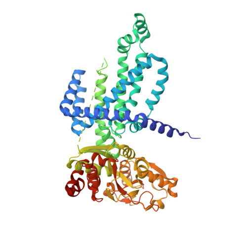
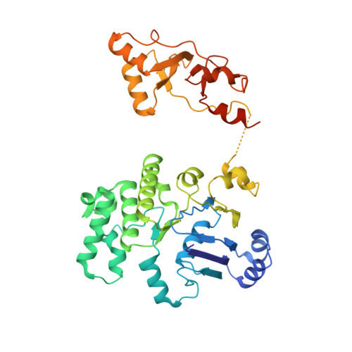
Gating by ionic strength and safety check by cyclic-di-AMP in the ABC transporter OpuA.

Sikkema, H.R.van den Noort, M.Rheinberger, J.de Boer, M.Krepel, S.T.Schuurman-Wolters, G.K.Paulino, C.Poolman, B.

(2020) Sci Adv 6

  • DOI: https://doi.org/10.1126/sciadv.abd7697
  • Primary Citation of Related Structures:  
    7AHC, 7AHD, 7AHE, 7AHH

  • PubMed Abstract: 

    (Micro)organisms are exposed to fluctuating environmental conditions, and adaptation to stress is essential for survival. Increased osmolality (hypertonicity) causes outflow of water and loss of turgor and is dangerous if the cell is not capable of rapidly restoring its volume. The osmoregulatory adenosine triphosphate-binding cassette transporter OpuA restores the cell volume by accumulating large amounts of compatible solute. OpuA is gated by ionic strength and inhibited by the second messenger cyclic-di-AMP, a molecule recently shown to affect many cellular processes. Despite the master regulatory role of cyclic-di-AMP, structural and functional insights into how the second messenger regulates (transport) proteins on the molecular level are lacking. Here, we present high-resolution cryo-electron microscopy structures of OpuA and in vitro activity assays that show how the osmoregulator OpuA is activated by high ionic strength and how cyclic-di-AMP acts as a backstop to prevent unbridled uptake of compatible solutes.


  • Organizational Affiliation

    Department of Biochemistry, Membrane Enzymology Group, Groningen Biomolecular Sciences and Biotechnology Institute and Zernike Institute for Advanced Materials, University of Groningen, Nijenborgh 4, 9747 AG, Groningen, Netherlands.


Macromolecules
Find similar proteins by:  (by identity cutoff)  |  3D Structure
Entity ID: 1
MoleculeChains Sequence LengthOrganismDetailsImage
ABC transporter permease subunit
A, B
585Lactococcus lactis subsp. lactisMutation(s): 0 
Gene Names: FEZ45_03205FIB48_03540GJI88_00475GJI90_00975LL275_1486N42_0725
Membrane Entity: Yes 
UniProt
Find proteins for A0A0V8ETW8 (Lactococcus lactis subsp. lactis)
Explore A0A0V8ETW8 
Go to UniProtKB:  A0A0V8ETW8
Entity Groups  
Sequence Clusters30% Identity50% Identity70% Identity90% Identity95% Identity100% Identity
UniProt GroupA0A0V8ETW8
Sequence Annotations
Expand
  • Reference Sequence
Find similar proteins by:  (by identity cutoff)  |  3D Structure
Entity ID: 2
MoleculeChains Sequence LengthOrganismDetailsImage
ABC-type proline/glycine betaine transport system ATPase component
C, D
408Lactococcus lactis subsp. lactisMutation(s): 0 
Gene Names: FEZ45_03200GJI88_00480GJI90_00980KF282_2364LL275_1487M20_1239N42_0726
UniProt
Find proteins for Q9KIF7 (Lactococcus lactis subsp. lactis (strain IL1403))
Explore Q9KIF7 
Go to UniProtKB:  Q9KIF7
Entity Groups  
Sequence Clusters30% Identity50% Identity70% Identity90% Identity95% Identity100% Identity
UniProt GroupQ9KIF7
Sequence Annotations
Expand
  • Reference Sequence
Small Molecules
Ligands 3 Unique
IDChains Name / Formula / InChI Key2D Diagram3D Interactions
2BA (Subject of Investigation/LOI)
Query on 2BA

Download Ideal Coordinates CCD File 
H [auth D](2R,3R,3aS,5R,7aR,9R,10R,10aS,12R,14aR)-2,9-bis(6-amino-9H-purin-9-yl)octahydro-2H,7H-difuro[3,2-d:3',2'-j][1,3,7,9,2,8 ]tetraoxadiphosphacyclododecine-3,5,10,12-tetrol 5,12-dioxide
C20 H24 N10 O12 P2
PDXMFTWFFKBFIN-XPWFQUROSA-N
ANP
Query on ANP

Download Ideal Coordinates CCD File 
F [auth C],
G [auth D]
PHOSPHOAMINOPHOSPHONIC ACID-ADENYLATE ESTER
C10 H17 N6 O12 P3
PVKSNHVPLWYQGJ-KQYNXXCUSA-N
BET
Query on BET

Download Ideal Coordinates CCD File 
E [auth A]TRIMETHYL GLYCINE
C5 H12 N O2
KWIUHFFTVRNATP-UHFFFAOYSA-O
Experimental Data & Validation

Experimental Data

  • Method: ELECTRON MICROSCOPY
  • Resolution: 3.50 Å
  • Aggregation State: PARTICLE 
  • Reconstruction Method: SINGLE PARTICLE 
EM Software:
TaskSoftware PackageVersion
MODEL REFINEMENTPHENIX
RECONSTRUCTIONRELION

Structure Validation

View Full Validation Report


Entry History & Funding Information

Deposition Data


Funding OrganizationLocationGrant Number
European Research Council (ERC)Netherlands670578
Netherlands Organisation for Scientific Research (NWO)Netherlands722.017.001
Netherlands Organisation for Scientific Research (NWO)Netherlands740.018.016

Revision History  (Full details and data files)

  • Version 1.0: 2020-11-25
    Type: Initial release
  • Version 1.1: 2020-12-02
    Changes: Database references
  • Version 2.0: 2023-11-15
    Changes: Atomic model, Data collection, Database references