METTL3 Inhibitors for Epitranscriptomic Modulation of Cellular Processes.
Moroz-Omori, E.V., Huang, D., Kumar Bedi, R., Cheriyamkunnel, S.J., Bochenkova, E., Dolbois, A., Rzeczkowski, M.D., Li, Y., Wiedmer, L., Caflisch, A.(2021) ChemMedChem 16: 3035-3043
- PubMed: 34237194 Search on PubMedSearch on PubMed Central
- DOI: https://doi.org/10.1002/cmdc.202100291
- Primary Citation Related Structures:
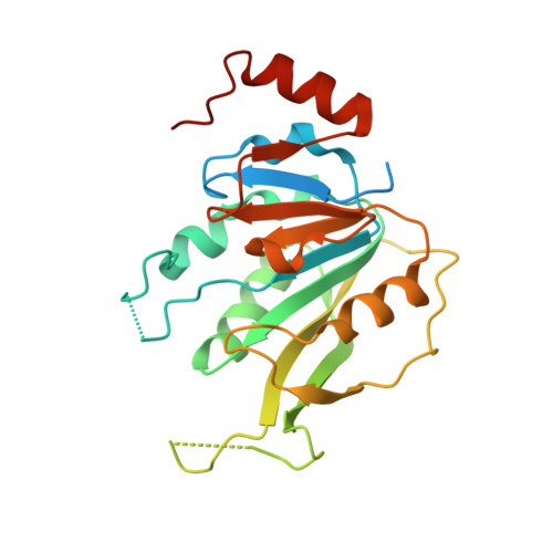
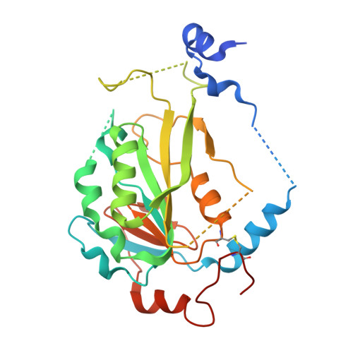
7ACD - PubMed Abstract:
The methylase METTL3 is the writer enzyme of the N 6 -methyladenosine (m 6 A) modification of RNA. Using a structure-based drug discovery approach, we identified a METTL3 inhibitor with potency in a biochemical assay of 280 nM, while its enantiomer is 100 times less active. We observed a dose-dependent reduction in the m 6 A methylation level of mRNA in several cell lines treated with the inhibitor already after 16 h of treatment, which lasted for at least 6 days. Importantly, the prolonged incubation (up to 6 days) with the METTL3 inhibitor did not alter levels of other RNA modifications (i. e., m 1 A, m 6 A m , m 7 G), suggesting selectivity of the developed compound towards other RNA methyltransferases.
- Department of Biochemistry, University of Zurich, Winterthurerstrasse 190, 8057, Zurich, Switzerland.
Organizational Affiliation: