7AAE

Crystal structure of Human serum albumin in complex with myristic acid at 2.27 Angstrom Resolution


Experimental Data Snapshot

  • Method: X-RAY DIFFRACTION
  • Resolution: 2.27 Å
  • R-Value Free: 0.291 
  • R-Value Work: 0.224 

wwPDB Validation   3D Report Full Report


Ligand Structure Quality Assessment 


This is version 1.2 of the entry. See complete history


Literature

Unveiling the binding mode of perfluorooctanoic acid to human serum albumin.

Maso, L.Trande, M.Liberi, S.Moro, G.Daems, E.Linciano, S.Sobott, F.Covaceuszach, S.Cassetta, A.Fasolato, S.Moretto, L.M.De Wael, K.Cendron, L.Angelini, A.

(2021) Protein Sci 30: 830-841

  • DOI: https://doi.org/10.1002/pro.4036
  • Primary Citation of Related Structures:  
    7AAE, 7AAI

  • PubMed Abstract: 

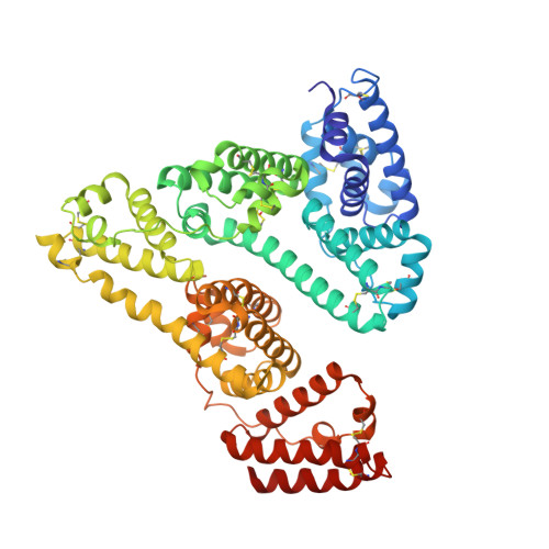
    Perfluorooctanoic acid (PFOA) is a toxic compound that is absorbed and distributed throughout the body by noncovalent binding to serum proteins such as human serum albumin (hSA). Though the interaction between PFOA and hSA has been already assessed using various analytical techniques, a high resolution and detailed analysis of the binding mode is still lacking. We report here the crystal structure of hSA in complex with PFOA and a medium-chain saturated fatty acid (FA). A total of eight distinct binding sites, four occupied by PFOAs and four by FAs, have been identified. In solution binding studies confirmed the 4:1 PFOA-hSA stoichiometry and revealed the presence of one high and three low affinity binding sites. Competition experiments with known hSA-binding drugs allowed locating the high affinity binding site in sub-domain IIIA. The elucidation of the molecular basis of the interaction between PFOA and hSA might provide not only a better assessment of the absorption and elimination mechanisms of these compounds in vivo but also have implications for the development of novel molecular receptors for diagnostic and biotechnological applications.


  • Organizational Affiliation

    Department of Biology, University of Padua, Padova, Italy.


Macromolecules
Find similar proteins by:  (by identity cutoff)  |  3D Structure
Entity ID: 1
MoleculeChains Sequence LengthOrganismDetailsImage
AlbuminA [auth AAA]584Homo sapiensMutation(s): 0 
Gene Names: ALBGIG20GIG42PRO0903PRO1708PRO2044PRO2619PRO2675UNQ696/PRO1341
UniProt & NIH Common Fund Data Resources
Find proteins for P02768 (Homo sapiens)
Explore P02768 
Go to UniProtKB:  P02768
PHAROS:  P02768
GTEx:  ENSG00000163631 
Entity Groups  
Sequence Clusters30% Identity50% Identity70% Identity90% Identity95% Identity100% Identity
UniProt GroupP02768
Sequence Annotations
Expand
  • Reference Sequence
Small Molecules
Ligands 4 Unique
IDChains Name / Formula / InChI Key2D Diagram3D Interactions
MYR (Subject of Investigation/LOI)
Query on MYR

Download Ideal Coordinates CCD File 
B [auth AAA]
C [auth AAA]
D [auth AAA]
E [auth AAA]
F [auth AAA]
B [auth AAA],
C [auth AAA],
D [auth AAA],
E [auth AAA],
F [auth AAA],
G [auth AAA]
MYRISTIC ACID
C14 H28 O2
TUNFSRHWOTWDNC-UHFFFAOYSA-N
MPD
Query on MPD

Download Ideal Coordinates CCD File 
H [auth AAA],
I [auth AAA],
J [auth AAA],
K [auth AAA],
L [auth AAA]
(4S)-2-METHYL-2,4-PENTANEDIOL
C6 H14 O2
SVTBMSDMJJWYQN-YFKPBYRVSA-N
PO4
Query on PO4

Download Ideal Coordinates CCD File 
N [auth AAA]PHOSPHATE ION
O4 P
NBIIXXVUZAFLBC-UHFFFAOYSA-K
FMT
Query on FMT

Download Ideal Coordinates CCD File 
M [auth AAA]FORMIC ACID
C H2 O2
BDAGIHXWWSANSR-UHFFFAOYSA-N
Experimental Data & Validation

Experimental Data

  • Method: X-RAY DIFFRACTION
  • Resolution: 2.27 Å
  • R-Value Free: 0.291 
  • R-Value Work: 0.224 
  • Space Group: I 1 2 1
Unit Cell:
Length ( Å )Angle ( ˚ )
a = 95.315α = 90
b = 38.537β = 104.45
c = 184.28γ = 90
Software Package:
Software NamePurpose
REFMACrefinement
xia2data reduction
Aimlessdata scaling
PHASERphasing
PHENIXrefinement

Structure Validation

View Full Validation Report


Ligand Structure Quality Assessment 


Entry History 

Deposition Data

Revision History  (Full details and data files)

  • Version 1.0: 2021-02-24
    Type: Initial release
  • Version 1.1: 2021-03-31
    Changes: Database references
  • Version 1.2: 2024-01-31
    Changes: Data collection, Database references, Derived calculations, Refinement description