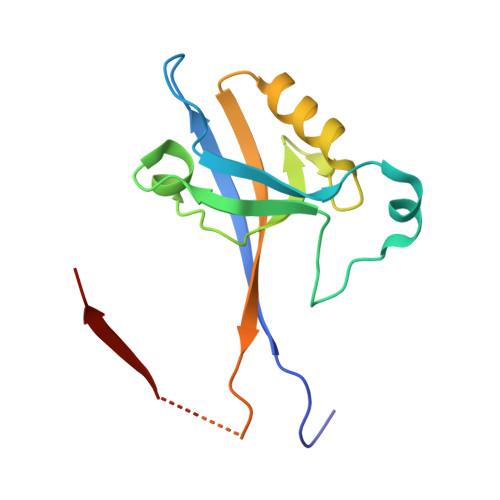
7A9B | pdb_00007a9b

Crystal structure of Shank1 PDZ domain with ARAP3-derived peptide


Experimental Data Snapshot

  • Method: X-RAY DIFFRACTION
  • Resolution: 2.00 Å
  • R-Value Free: 
    0.249 (Depositor), 0.251 (DCC) 
  • R-Value Work: 
    0.201 (Depositor), 0.203 (DCC) 
  • R-Value Observed: 
    0.203 (Depositor) 

Starting Model: experimental
View more details

wwPDB Validation 3D Report Full Report

Validation slider image for 7A9B

This is version 1.2 of the entry. See complete history. 

Literature

Integrated analysis of Shank1 PDZ interactions with C-terminal and internal binding motifs.

Ali, M., McAuley, M.M., Luchow, S., Knapp, S., Joerger, A.C., Ivarsson, Y.

(2021) Curr Res Struct Biol 3: 41-50

  • DOI: https://doi.org/10.1016/j.crstbi.2021.01.001
  • Primary Citation Related Structures: 
    7A9B

  • PubMed Abstract: 

    PDZ domains constitute a large family of modular domains that are well-known for binding C-terminal motifs of target proteins. Some of them also bind to internal PDZ binding motifs (PDZbms), but this aspect of the PDZ interactome is poorly studied. Here we explored internal PDZbm-mediated interactions using the PDZ domain of Shank1 as a model. We identified a series of human Shank1 ligands with C-terminal or internal PDZbms using proteomic peptide-phage display, and established that while the consensus sequence of C-terminal ligands is x-T-x-(L/F)-COOH, the consensus of internal PDZbm is exclusively x-T-x-F-x, where x is any amino acid. We found that the affinities of PDZbm interactions are in the low micromolar range. The crystal structure of the complex between Shank1 PDZ and an internal PDZbm revealed that the binding mode of internal PDZbms was similar to that of C-terminal ligands. Pull-down experiments confirmed that both C-terminal and internal PDZbm interactions can occur in the context of full-length proteins. Our study expands the interactome of Shank1 and hints at a largely unexplored interaction space of PDZ domains.


  • Organizational Affiliation: 
    • Department of Chemistry - BMC, Uppsala University, Husargatan 3, 751 23, Uppsala, Sweden.

Macromolecule Content 

  • Total Structure Weight: 30.18 kDa 
  • Atom Count: 1,992 
  • Modeled Residue Count: 253 
  • Deposited Residue Count: 280 
  • Unique protein chains: 1

Macromolecules

Find similar proteins by:|  3D Structure
Entity ID: 1
MoleculeChains  Sequence LengthOrganismDetailsImage
SH3 and multiple ankyrin repeat domains protein 1,Arf-GAP with Rho-GAP domain, ANK repeat and PH domain-containing protein 3
A, B
140Rattus norvegicus, Homo sapiens
This entity is chimeric
Mutation(s): 0 
Gene Names: Shank1, ARAP3, CENTD3
UniProt & NIH Common Fund Data Resources
Find proteins for Q8WWN8 (Homo sapiens)
Explore Q8WWN8 
Go to UniProtKB:  Q8WWN8
PHAROS:  Q8WWN8
GTEx:  ENSG00000120318 
Find proteins for Q9WV48 (Rattus norvegicus)
Explore Q9WV48 
Go to UniProtKB:  Q9WV48
Entity Groups
Sequence Clusters30% Identity50% Identity70% Identity90% Identity95% Identity100% Identity
UniProt GroupsQ9WV48Q8WWN8
Sequence Annotations
Expand
Reference Sequence

Small Molecules

Ligands 1 Unique
IDChains Name / Formula / InChI Key2D Diagram3D Interactions
EDO

Query on EDO


Download:Ideal Coordinates CCD File
C [auth B]1,2-ETHANEDIOL
C2 H6 O2
LYCAIKOWRPUZTN-UHFFFAOYSA-N

Experimental Data & Validation

Experimental Data

  • Method: X-RAY DIFFRACTION
  • Resolution: 2.00 Å
  • R-Value Free:  0.249 (Depositor), 0.251 (DCC) 
  • R-Value Work:  0.201 (Depositor), 0.203 (DCC) 
  • R-Value Observed: 0.203 (Depositor) 
Space Group: P 61 2 2
Unit Cell:
Length ( Å )Angle ( ˚ )
a = 67.799α = 90
b = 67.799β = 90
c = 248.967γ = 120
Software Package:
Software NamePurpose
PHENIXrefinement
XDSdata reduction
Aimlessdata scaling
PHASERphasing

Structure Validation

View Full Validation Report


Entry History 

& Funding Information

Deposition Data


Funding OrganizationLocationGrant Number
H2020 Marie Curie Actions of the European Commission675341

Revision History  (Full details and data files)

  • Version 1.0: 2020-10-21
    Type: Initial release
  • Version 1.1: 2024-01-31
    Changes: Data collection, Database references, Refinement description
  • Version 1.2: 2026-09-30
    Changes: Database references, Structure summary