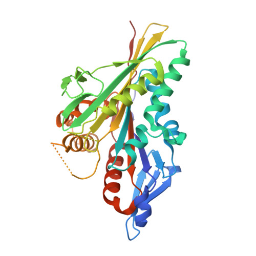
Structural snapshots of the kinesin-2 OSM-3 along its nucleotide cycle: implications for the ATP hydrolysis mechanism.
Varela, P.F., Chenon, M., Velours, C., Verhey, K.J., Menetrey, J., Gigant, B.(2021) FEBS Open Bio 11: 564-577
- PubMed: 33513284
- DOI: https://doi.org/10.1002/2211-5463.13101
- Primary Citation of Related Structures:
7A3Z, 7A40, 7A5E - PubMed Abstract:
Motile kinesins are motor proteins that translocate along microtubules as they hydrolyze ATP. They share a conserved motor domain which harbors both ATPase and microtubule-binding activities. An ATP hydrolysis mechanism involving two water molecules has been proposed based on the structure of the kinesin-5 Eg5 bound to an ATP analog. Whether this mechanism is general in the kinesin superfamily remains uncertain. Here, we present structural snapshots of the motor domain of OSM-3 along its nucleotide cycle. OSM-3 belongs to the homodimeric kinesin-2 subfamily and is the Caenorhabditis elegans homologue of human KIF17. OSM-3 bound to ADP or devoid of a nucleotide shows features of ADP-kinesins with a docked neck linker. When bound to an ATP analog, OSM-3 adopts a conformation similar to those of several ATP-like kinesins, either isolated or bound to tubulin. Moreover, the OSM-3 nucleotide-binding site is virtually identical to that of ATP-like Eg5, demonstrating a shared ATPase mechanism. Therefore, our data extend to kinesin-2 the two-water ATP hydrolysis mechanism and further suggest that it is universal within the kinesin superfamily. PROTEIN DATABASE ENTRIES: 7A3Z, 7A40, 7A5E.
- Université Paris-Saclay, CEA, CNRS, Institute for Integrative Biology of the Cell (I2BC), Gif-sur-Yvette, France.
Organizational Affiliation:


