6ZQR

Crystal structure of tetrameric fibrinogen-like recognition domain of FIBCD1 with GlcNAc ligand bound


Experimental Data Snapshot

  • Method: X-RAY DIFFRACTION
  • Resolution: 1.93 Å
  • R-Value Free: 0.191 
  • R-Value Work: 0.167 
  • R-Value Observed: 0.168 

wwPDB Validation   3D Report Full Report


Ligand Structure Quality Assessment 


This is version 1.3 of the entry. See complete history


Literature

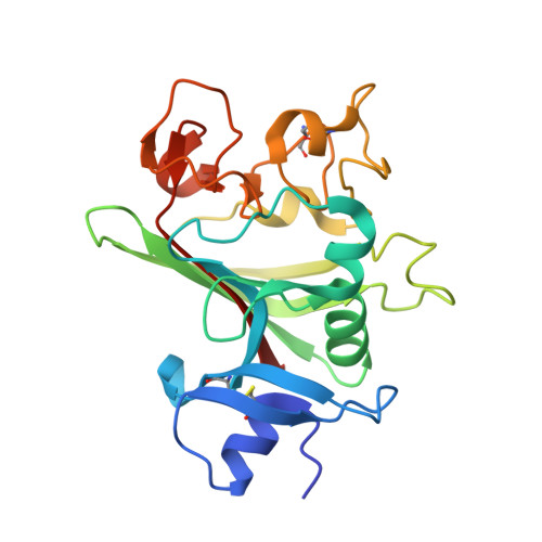
Crystal structures of human immune protein FIBCD1 suggest an extended binding site compatible with recognition of pathogen associated carbohydrate motifs

Williams, H.M.Moeller, J.B.Burns, I.Schlosser, A.Sorensen, G.L.Greenhough, T.J.Holmskov, U.Shrive, A.K.

(2023) J Biol Chem 


Macromolecules
Find similar proteins by:  (by identity cutoff)  |  3D Structure
Entity ID: 1
MoleculeChains Sequence LengthOrganismDetailsImage
Fibrinogen C domain-containing protein 1
A, B
226Homo sapiensMutation(s): 0 
Gene Names: FIBCD1UNQ701/PRO1346
UniProt & NIH Common Fund Data Resources
Find proteins for Q8N539 (Homo sapiens)
Explore Q8N539 
Go to UniProtKB:  Q8N539
PHAROS:  Q8N539
GTEx:  ENSG00000130720 
Entity Groups  
Sequence Clusters30% Identity50% Identity70% Identity90% Identity95% Identity100% Identity
UniProt GroupQ8N539
Sequence Annotations
Expand
  • Reference Sequence
Oligosaccharides

Help

Entity ID: 2
MoleculeChains Length2D Diagram Glycosylation3D Interactions
alpha-L-fucopyranose-(1-3)-[2-acetamido-2-deoxy-beta-D-glucopyranose-(1-4)][alpha-L-fucopyranose-(1-6)]2-acetamido-2-deoxy-beta-D-glucopyranose
C
4N-Glycosylation
Glycosylation Resources
GlyTouCan:  G63564LA
GlyCosmos:  G63564LA
Small Molecules
Ligands 4 Unique
IDChains Name / Formula / InChI Key2D Diagram3D Interactions
NAG (Subject of Investigation/LOI)
Query on NAG

Download Ideal Coordinates CCD File 
H [auth A]2-acetamido-2-deoxy-beta-D-glucopyranose
C8 H15 N O6
OVRNDRQMDRJTHS-FMDGEEDCSA-N
SO4
Query on SO4

Download Ideal Coordinates CCD File 
E [auth A],
F [auth A],
J [auth B],
K [auth B]
SULFATE ION
O4 S
QAOWNCQODCNURD-UHFFFAOYSA-L
ACY
Query on ACY

Download Ideal Coordinates CCD File 
G [auth A],
L [auth B],
M [auth B]
ACETIC ACID
C2 H4 O2
QTBSBXVTEAMEQO-UHFFFAOYSA-N
CA
Query on CA

Download Ideal Coordinates CCD File 
D [auth A],
I [auth B]
CALCIUM ION
Ca
BHPQYMZQTOCNFJ-UHFFFAOYSA-N
Experimental Data & Validation

Experimental Data

  • Method: X-RAY DIFFRACTION
  • Resolution: 1.93 Å
  • R-Value Free: 0.191 
  • R-Value Work: 0.167 
  • R-Value Observed: 0.168 
  • Space Group: P 4
  • Diffraction Data: https://doi.org/10.21252/r2nx-0425
Unit Cell:
Length ( Å )Angle ( ˚ )
a = 113.6α = 90
b = 113.6β = 90
c = 44.08γ = 90
Software Package:
Software NamePurpose
MOSFLMdata reduction
Aimlessdata scaling
MOLREPphasing
REFMACrefinement
PDB_EXTRACTdata extraction

Structure Validation

View Full Validation Report


Ligand Structure Quality Assessment 


Entry History & Funding Information

Deposition Data


Funding OrganizationLocationGrant Number
Science and Technology Funding CouncilUnited KingdomMX6388

Revision History  (Full details and data files)

  • Version 1.0: 2021-07-21
    Type: Initial release
  • Version 1.1: 2023-12-27
    Changes: Data collection, Database references
  • Version 1.2: 2024-01-10
    Changes: Database references
  • Version 1.3: 2024-01-31
    Changes: Refinement description