6ZMD

Crystal structure of HYPE covalently tethered to BiP bound to AMP-PNP


Experimental Data Snapshot

  • Method: X-RAY DIFFRACTION
  • Resolution: 2.64 Å
  • R-Value Free: 0.235 
  • R-Value Work: 0.192 
  • R-Value Observed: 0.194 

wwPDB Validation   3D Report Full Report


This is version 1.2 of the entry. See complete history


Literature

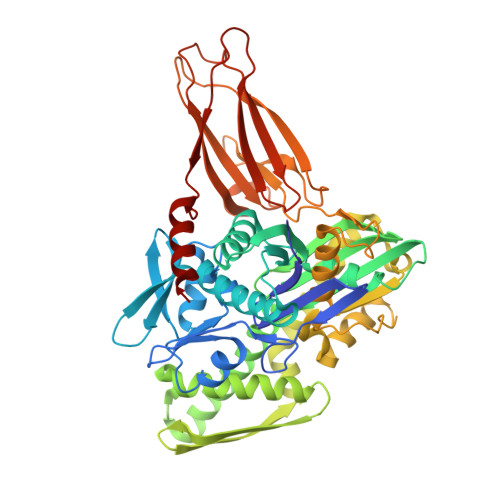
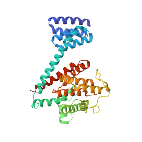
Specificity of AMPylation of the human chaperone BiP is mediated by TPR motifs of FICD.

Fauser, J.Gulen, B.Pogenberg, V.Pett, C.Pourjafar-Dehkordi, D.Krisp, C.Hopfner, D.Konig, G.Schluter, H.Feige, M.J.Zacharias, M.Hedberg, C.Itzen, A.

(2021) Nat Commun 12: 2426-2426

  • DOI: https://doi.org/10.1038/s41467-021-22596-0
  • Primary Citation of Related Structures:  
    6ZMD

  • PubMed Abstract: 

    To adapt to fluctuating protein folding loads in the endoplasmic reticulum (ER), the Hsp70 chaperone BiP is reversibly modified with adenosine monophosphate (AMP) by the ER-resident Fic-enzyme FICD/HYPE. The structural basis for BiP binding and AMPylation by FICD has remained elusive due to the transient nature of the enzyme-substrate-complex. Here, we use thiol-reactive derivatives of the cosubstrate adenosine triphosphate (ATP) to covalently stabilize the transient FICD:BiP complex and determine its crystal structure. The complex reveals that the TPR-motifs of FICD bind specifically to the conserved hydrophobic linker of BiP and thus mediate specificity for the domain-docked conformation of BiP. Furthermore, we show that both AMPylation and deAMPylation of BiP are not directly regulated by the presence of unfolded proteins. Together, combining chemical biology, crystallography and biochemistry, our study provides structural insights into a key regulatory mechanism that safeguards ER homeostasis.


  • Organizational Affiliation

    Department of Biochemistry and Signal Transduction, University Medical Center Hamburg-Eppendorf (UKE), Hamburg, Germany.


Macromolecules
Find similar proteins by:  (by identity cutoff)  |  3D Structure
Entity ID: 1
MoleculeChains Sequence LengthOrganismDetailsImage
Endoplasmic reticulum chaperone BiP527Homo sapiensMutation(s): 1 
Gene Names: HSPA5GRP78
EC: 3.6.4.10
UniProt & NIH Common Fund Data Resources
Find proteins for P11021 (Homo sapiens)
Explore P11021 
Go to UniProtKB:  P11021
PHAROS:  P11021
GTEx:  ENSG00000044574 
Entity Groups  
Sequence Clusters30% Identity50% Identity70% Identity90% Identity95% Identity100% Identity
UniProt GroupP11021
Sequence Annotations
Expand
  • Reference Sequence
Find similar proteins by:  (by identity cutoff)  |  3D Structure
Entity ID: 2
MoleculeChains Sequence LengthOrganismDetailsImage
Protein adenylyltransferase FICD344Homo sapiensMutation(s): 5 
Gene Names: FICDHIP13HYPEUNQ3041/PRO9857
EC: 2.7.7 (PDB Primary Data), 3.1.4 (PDB Primary Data)
UniProt & NIH Common Fund Data Resources
Find proteins for Q9BVA6 (Homo sapiens)
Explore Q9BVA6 
Go to UniProtKB:  Q9BVA6
PHAROS:  Q9BVA6
GTEx:  ENSG00000198855 
Entity Groups  
Sequence Clusters30% Identity50% Identity70% Identity90% Identity95% Identity100% Identity
UniProt GroupQ9BVA6
Sequence Annotations
Expand
  • Reference Sequence
Small Molecules
Ligands 4 Unique
IDChains Name / Formula / InChI Key2D Diagram3D Interactions
ANP
Query on ANP

Download Ideal Coordinates CCD File 
C [auth A]PHOSPHOAMINOPHOSPHONIC ACID-ADENYLATE ESTER
C10 H17 N6 O12 P3
PVKSNHVPLWYQGJ-KQYNXXCUSA-N
QMK (Subject of Investigation/LOI)
Query on QMK

Download Ideal Coordinates CCD File 
D [auth A]~{N}-[2-[1-[(2~{R},3~{R},4~{S},5~{R})-3,4-bis(oxidanyl)-5-[[tris(oxidanyl)-$l^{5}-phosphanyl]oxymethyl]oxolan-2-yl]-1,2,3-triazol-4-yl]ethyl]ethanamide
C11 H19 N4 O8 P
KBIJUYSCKWPFAX-GWOFURMSSA-N
PO4
Query on PO4

Download Ideal Coordinates CCD File 
G [auth A],
H [auth B]
PHOSPHATE ION
O4 P
NBIIXXVUZAFLBC-UHFFFAOYSA-K
MG
Query on MG

Download Ideal Coordinates CCD File 
E [auth A],
F [auth A]
MAGNESIUM ION
Mg
JLVVSXFLKOJNIY-UHFFFAOYSA-N
Experimental Data & Validation

Experimental Data

  • Method: X-RAY DIFFRACTION
  • Resolution: 2.64 Å
  • R-Value Free: 0.235 
  • R-Value Work: 0.192 
  • R-Value Observed: 0.194 
  • Space Group: P 41
Unit Cell:
Length ( Å )Angle ( ˚ )
a = 83.466α = 90
b = 83.466β = 90
c = 169.482γ = 90
Software Package:
Software NamePurpose
PHENIXrefinement
DIALSdata reduction
Aimlessdata scaling
MoRDaphasing

Structure Validation

View Full Validation Report


Entry History & Funding Information

Deposition Data


Funding OrganizationLocationGrant Number
German Research Foundation (DFG)GermanySFB1035 B05
Knut and Alice Wallenberg FoundationSweden--

Revision History  (Full details and data files)

  • Version 1.0: 2021-04-14
    Type: Initial release
  • Version 1.1: 2021-10-06
    Changes: Data collection, Database references, Structure summary
  • Version 1.2: 2024-01-31
    Changes: Data collection, Refinement description