6ZH9 | pdb_00006zh9

Ternary complex CR3022 H11-H4 and RBD (SARS-CoV-2)


Experimental Data Snapshot

  • Method: X-RAY DIFFRACTION
  • Resolution: 3.31 Å
  • R-Value Free: 
    0.305 (Depositor), 0.313 (DCC) 
  • R-Value Work: 
    0.261 (Depositor), 0.265 (DCC) 

Starting Model: experimental
View more details

wwPDB Validation 3D Report Full Report

Validation slider image for 6ZH9

This is version 1.3 of the entry. See complete history

Literature

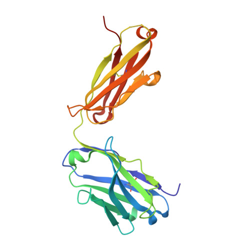
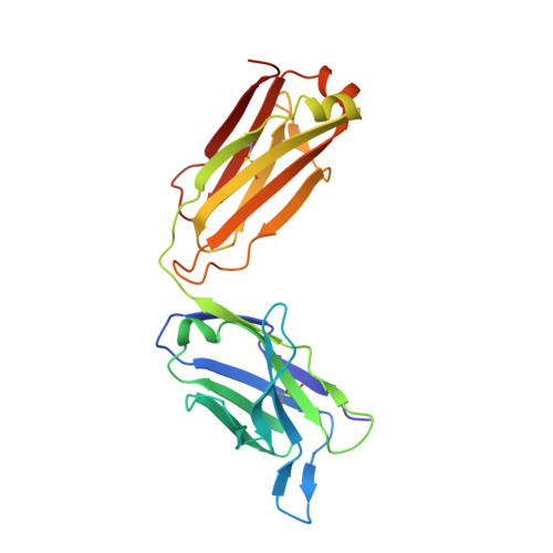
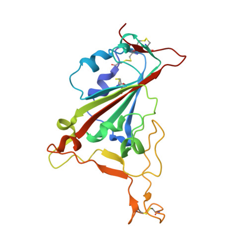
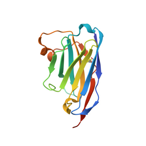
Neutralizing nanobodies bind SARS-CoV-2 spike RBD and block interaction with ACE2.

Huo, J.Le Bas, A.Ruza, R.R.Duyvesteyn, H.M.E.Mikolajek, H.Malinauskas, T.Tan, T.K.Rijal, P.Dumoux, M.Ward, P.N.Ren, J.Zhou, D.Harrison, P.J.Weckener, M.Clare, D.K.Vogirala, V.K.Radecke, J.Moynie, L.Zhao, Y.Gilbert-Jaramillo, J.Knight, M.L.Tree, J.A.Buttigieg, K.R.Coombes, N.Elmore, M.J.Carroll, M.W.Carrique, L.Shah, P.N.M.James, W.Townsend, A.R.Stuart, D.I.Owens, R.J.Naismith, J.H.

(2020) Nat Struct Mol Biol 27: 846-854

  • DOI: https://doi.org/10.1038/s41594-020-0469-6
  • Primary Citation Related Structures: 
    6ZH9

  • PubMed Abstract: 

    The SARS-CoV-2 virus is more transmissible than previous coronaviruses and causes a more serious illness than influenza. The SARS-CoV-2 receptor binding domain (RBD) of the spike protein binds to the human angiotensin-converting enzyme 2 (ACE2) receptor as a prelude to viral entry into the cell. Using a naive llama single-domain antibody library and PCR-based maturation, we have produced two closely related nanobodies, H11-D4 and H11-H4, that bind RBD (K D of 39 and 12 nM, respectively) and block its interaction with ACE2. Single-particle cryo-EM revealed that both nanobodies bind to all three RBDs in the spike trimer. Crystal structures of each nanobody-RBD complex revealed how both nanobodies recognize the same epitope, which partly overlaps with the ACE2 binding surface, explaining the blocking of the RBD-ACE2 interaction. Nanobody-Fc fusions showed neutralizing activity against SARS-CoV-2 (4-6 nM for H11-H4, 18 nM for H11-D4) and additive neutralization with the SARS-CoV-1/2 antibody CR3022.


  • Organizational Affiliation
    • Structural Biology, The Rosalind Franklin Institute, Harwell Science & Innovation Campus, Didcot, UK.

Macromolecule Content 

  • Total Structure Weight: 84.22 kDa 
  • Atom Count: 5,906 
  • Modeled Residue Count: 758 
  • Deposited Residue Count: 766 
  • Unique protein chains: 4

Macromolecules

Find similar proteins by:|  3D Structure
Entity ID: 1
MoleculeChains  Sequence LengthOrganismDetailsImage
CR3022 heavyA [auth HHH]216Homo sapiensMutation(s): 0 
Find similar proteins by:|  3D Structure
Entity ID: 2
MoleculeChains  Sequence LengthOrganismDetailsImage
CR3022 Light chainB [auth LLL]219Homo sapiensMutation(s): 0 
Find similar proteins by:|  3D Structure
Entity ID: 3
MoleculeChains  Sequence LengthOrganismDetailsImage
Spike glycoproteinC [auth EEE]197Severe acute respiratory syndrome coronavirus 2Mutation(s): 0 
Gene Names: S2
UniProt
Find proteins for P0DTC2 (Severe acute respiratory syndrome coronavirus 2)
Explore P0DTC2 
Go to UniProtKB:  P0DTC2
Entity Groups
Sequence Clusters30% Identity50% Identity70% Identity90% Identity95% Identity100% Identity
UniProt GroupP0DTC2
Sequence Annotations
Expand
Reference Sequence
Find similar proteins by:|  3D Structure
Entity ID: 4
MoleculeChains  Sequence LengthOrganismDetailsImage
Nanobody H11-H4D [auth FFF]134Lama glamaMutation(s): 0 

Experimental Data & Validation

Experimental Data

  • Method: X-RAY DIFFRACTION
  • Resolution: 3.31 Å
  • R-Value Free:  0.305 (Depositor), 0.313 (DCC) 
  • R-Value Work:  0.261 (Depositor), 0.265 (DCC) 
Space Group: P 42 21 2
Unit Cell:
Length ( Å )Angle ( ˚ )
a = 156.411α = 90
b = 156.411β = 90
c = 116.205γ = 90
Software Package:
Software NamePurpose
REFMACrefinement
XDSdata reduction
pointlessdata scaling
PHASERphasing

Structure Validation

View Full Validation Report


Entry History 

& Funding Information

Deposition Data


Funding OrganizationLocationGrant Number
Wellcome TrustUnited Kingdom100209/Z/12/Z).
Engineering and Physical Sciences Research CouncilUnited KingdomRFI

Revision History  (Full details and data files)

  • Version 1.0: 2020-09-02
    Type: Initial release
  • Version 1.1: 2020-09-16
    Changes: Database references
  • Version 1.2: 2024-01-24
    Changes: Data collection, Database references, Derived calculations, Refinement description
  • Version 1.3: 2024-11-20
    Changes: Structure summary