6Z9F

1.56 A structure of human apoferritin obtained from data subset of Titan Mono-BCOR microscope


Experimental Data Snapshot

  • Method: ELECTRON MICROSCOPY
  • Resolution: 1.56 Å
  • Aggregation State: PARTICLE 
  • Reconstruction Method: SINGLE PARTICLE 

wwPDB Validation   3D Report Full Report


This is version 1.1 of the entry. See complete history


Literature

Atomic-resolution protein structure determination by cryo-EM.

Yip, K.M.Fischer, N.Paknia, E.Chari, A.Stark, H.

(2020) Nature 587: 157-161

  • DOI: https://doi.org/10.1038/s41586-020-2833-4
  • Primary Citation of Related Structures:  
    6Z6U, 6Z9E, 6Z9F, 7A6A, 7A6B

  • PubMed Abstract: 

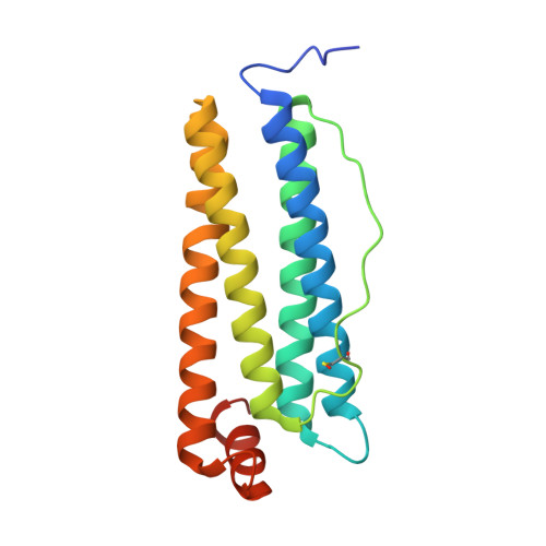
    Single-particle electron cryo-microscopy (cryo-EM) is a powerful method for solving the three-dimensional structures of biological macromolecules. The technological development of transmission electron microscopes, detectors and automated procedures in combination with user-friendly image processing software and ever-increasing computational power have made cryo-EM a successful and expanding technology over the past decade 1 . At resolutions better than 4 Å, atomic model building starts to become possible, but the direct visualization of true atomic positions in protein structure determination requires much higher (better than 1.5 Å) resolution, which so far has not been attained by cryo-EM. The direct visualization of atom positions is essential for understanding the mechanisms of protein-catalysed chemical reactions, and for studying how drugs bind to and interfere with the function of proteins 2 . Here we report a 1.25 Å-resolution structure of apoferritin obtained by cryo-EM with a newly developed electron microscope that provides, to our knowledge, unprecedented structural detail. Our apoferritin structure has almost twice the 3D information content of the current world record reconstruction (at 1.54 Å resolution 3 ). We can visualize individual atoms in a protein, see density for hydrogen atoms and image single-atom chemical modifications. Beyond the nominal improvement in resolution, we also achieve a substantial improvement in the quality of the cryo-EM density map, which is highly relevant for using cryo-EM in structure-based drug design.


  • Organizational Affiliation

    Department of Structural Dynamics, MPI for Biophysical Chemistry, Göttingen, Germany.


Macromolecules
Find similar proteins by:  (by identity cutoff)  |  3D Structure
Entity ID: 1
MoleculeChains Sequence LengthOrganismDetailsImage
Ferritin heavy chain183Homo sapiensMutation(s): 0 
Gene Names: FTH1FTHFTHL6OK/SW-cl.84PIG15
EC: 1.16.3.1
UniProt & NIH Common Fund Data Resources
Find proteins for P02794 (Homo sapiens)
Explore P02794 
Go to UniProtKB:  P02794
PHAROS:  P02794
GTEx:  ENSG00000167996 
Entity Groups  
Sequence Clusters30% Identity50% Identity70% Identity90% Identity95% Identity100% Identity
UniProt GroupP02794
Sequence Annotations
Expand
  • Reference Sequence
Small Molecules
Ligands 1 Unique
IDChains Name / Formula / InChI Key2D Diagram3D Interactions
NA
Query on NA

Download Ideal Coordinates CCD File 
AA [auth 1]
AB [auth P]
BA [auth 1]
BB [auth X]
CA [auth K]
AA [auth 1],
AB [auth P],
BA [auth 1],
BB [auth X],
CA [auth K],
CB [auth 6],
DA [auth a],
DB [auth 6],
EA [auth B],
FA [auth B],
GA [auth E],
HA [auth e],
IA [auth r],
JA [auth G],
KA [auth I],
LA [auth M],
MA [auth M],
NA [auth O],
OA [auth Q],
PA [auth S],
QA [auth U],
RA [auth W],
SA [auth Y],
TA [auth 2],
UA [auth 2],
VA [auth 4],
WA [auth 4],
XA [auth F],
Y [auth A],
YA [auth H],
Z [auth A],
ZA [auth H]
SODIUM ION
Na
FKNQFGJONOIPTF-UHFFFAOYSA-N
Modified Residues  1 Unique
IDChains TypeFormula2D DiagramParent
CSX
Query on CSX
A
B [auth 1]
C [auth K]
D [auth a]
E [auth B]
A,
B [auth 1],
C [auth K],
D [auth a],
E [auth B],
F [auth E],
G [auth e],
H [auth r],
I [auth G],
J [auth I],
K [auth M],
L [auth O],
M [auth Q],
N [auth S],
O [auth U],
P [auth W],
Q [auth Y],
R [auth 2],
S [auth 4],
T [auth F],
U [auth H],
V [auth P],
W [auth X],
X [auth 6]
L-PEPTIDE LINKINGC3 H7 N O3 SCYS
Experimental Data & Validation

Experimental Data

  • Method: ELECTRON MICROSCOPY
  • Resolution: 1.56 Å
  • Aggregation State: PARTICLE 
  • Reconstruction Method: SINGLE PARTICLE 
EM Software:
TaskSoftware PackageVersion
MODEL REFINEMENTREFMAC
RECONSTRUCTIONCow

Structure Validation

View Full Validation Report


Entry History & Funding Information

Deposition Data


Funding OrganizationLocationGrant Number
German Research Foundation (DFG)Germany--
Max Planck SocietyGermany--

Revision History  (Full details and data files)

  • Version 1.0: 2020-06-24
    Type: Initial release
  • Version 1.1: 2021-02-10
    Changes: Database references, Derived calculations