6YK4

Structure of the AMPA receptor GluA2o ligand-binding domain (S1S2J) in complex with the compound ( S) - 1- [2'-Amino-2'-carboxyethyl]-6-methyl-5 ,7- dihydropyrrolo[3,4-d]pyrimidin-2,4(1H,3H)-dione at resolution 1.00A


Experimental Data Snapshot

  • Method: X-RAY DIFFRACTION
  • Resolution: 1.00 Å
  • R-Value Free: 0.120 
  • R-Value Work: 0.106 
  • R-Value Observed: 0.107 

wwPDB Validation   3D Report Full Report


Ligand Structure Quality Assessment 


This is version 1.3 of the entry. See complete history


Literature

Ionotropic Glutamate Receptor GluA2 in Complex with Bicyclic Pyrimidinedione-Based Compounds: When Small Compound Modifications Have Distinct Effects on Binding Interactions.

Frydenvang, K.Pickering, D.S.Kshirsagar, G.U.Chemi, G.Gemma, S.Sprogoe, D.Kærn, A.M.Brogi, S.Campiani, G.Butini, S.Kastrup, J.S.

(2020) ACS Chem Neurosci 11: 1791-1800

  • DOI: https://doi.org/10.1021/acschemneuro.0c00195
  • Primary Citation of Related Structures:  
    6YK2, 6YK3, 6YK4, 6YK5, 6YK6

  • PubMed Abstract: 

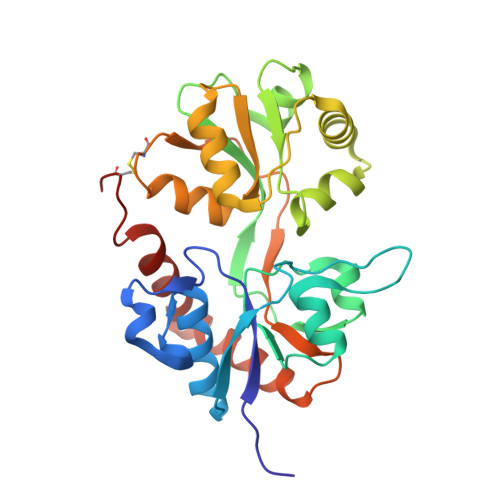
    ( S )-2-Amino-3-(5-methyl-3-hydroxyisoxazol-4-yl)propanoic acid (AMPA) receptors comprise an important class of ionotropic glutamate receptors activated by glutamate in the central nervous system. These receptors have been shown to be involved in brain diseases, for example, Alzheimer's disease and epilepsy. To understand the functional role of AMPA receptors at the molecular level and their potential as targets for drugs, development of tool compounds is essential. We have previously reported the synthesis of six bicyclic pyrimidinedione-based analogues of willardiine with differences limited to the pyrimidinedione-fused five-membered rings. Despite minor molecular differences, we observed >500-fold difference in binding affinity of the compounds at full-length GluA2. Here, we report binding affinities and the binding mode of these compounds at the ligand-binding domain of GluA2 using X-ray crystallography. The structures revealed similar binding modes, with distinct differences in the interaction between GluA2 and the compounds. The methylene ( 2 ) and sulfur ( 3 ) containing compounds showed the greatest binding affinities. Changing the dihydrothiophene ( 3 ) into pyrrolidine ( 4 ), N -methyl pyrrolidine ( 5 ), or dihydrofuran ( 6 ) induced flexibility in the position of a binding-site water molecule and changes in the hydrogen-bonding network between compound, water, and GluA2. This might be essential for explaining the reduced binding affinity of these compounds. The weakest binding affinity was observed when the aliphatic oxygen containing dihydrofuran ( 6 ) was changed into an aromatic furan system ( 7 ). Molecular docking studies revealed two possible orientations of 7 , whereas only one binding mode was observed for the other analogues. This could likely contribute to the weakest binding affinity of 7 at GluA2.


  • Organizational Affiliation

    Department of Drug Design and Pharmacology, University of Copenhagen, Jagtvej 162, DK-2100 Copenhagen, Denmark.


Macromolecules
Find similar proteins by:  (by identity cutoff)  |  3D Structure
Entity ID: 1
MoleculeChains Sequence LengthOrganismDetailsImage
Glutamate receptor 2,Glutamate receptor 2264Rattus norvegicusMutation(s): 0 
Gene Names: Gria2Glur2
UniProt
Find proteins for P19491 (Rattus norvegicus)
Explore P19491 
Go to UniProtKB:  P19491
Entity Groups  
Sequence Clusters30% Identity50% Identity70% Identity90% Identity95% Identity100% Identity
UniProt GroupP19491
Sequence Annotations
Expand
  • Reference Sequence
Small Molecules
Ligands 5 Unique
IDChains Name / Formula / InChI Key2D Diagram3D Interactions
CG8 (Subject of Investigation/LOI)
Query on CG8

Download Ideal Coordinates CCD File 
K [auth A](2~{S})-2-azanyl-3-[6-methyl-2,4-bis(oxidanylidene)-5,7-dihydropyrrolo[3,4-d]pyrimidin-1-yl]propanoic acid
C10 H14 N4 O4
BJXZWHLUHLDGNK-LURJTMIESA-N
SO4
Query on SO4

Download Ideal Coordinates CCD File 
B [auth A],
C [auth A],
D [auth A],
E [auth A]
SULFATE ION
O4 S
QAOWNCQODCNURD-UHFFFAOYSA-L
GOL
Query on GOL

Download Ideal Coordinates CCD File 
F [auth A],
G [auth A],
H [auth A],
I [auth A]
GLYCEROL
C3 H8 O3
PEDCQBHIVMGVHV-UHFFFAOYSA-N
CL
Query on CL

Download Ideal Coordinates CCD File 
L [auth A],
M [auth A]
CHLORIDE ION
Cl
VEXZGXHMUGYJMC-UHFFFAOYSA-M
LI
Query on LI

Download Ideal Coordinates CCD File 
J [auth A]LITHIUM ION
Li
HBBGRARXTFLTSG-UHFFFAOYSA-N
Binding Affinity Annotations 
IDSourceBinding Affinity
CG8 Binding MOAD:  6YK4 Ki: 7890 (nM) from 1 assay(s)
Experimental Data & Validation

Experimental Data

  • Method: X-RAY DIFFRACTION
  • Resolution: 1.00 Å
  • R-Value Free: 0.120 
  • R-Value Work: 0.106 
  • R-Value Observed: 0.107 
  • Space Group: P 21 21 2
Unit Cell:
Length ( Å )Angle ( ˚ )
a = 59.213α = 90
b = 96.542β = 90
c = 48.543γ = 90
Software Package:
Software NamePurpose
SCALAdata scaling
PHENIXrefinement
PDB_EXTRACTdata extraction
XDSdata reduction
PHASERphasing

Structure Validation

View Full Validation Report


Ligand Structure Quality Assessment 


Entry History 

Deposition Data

Revision History  (Full details and data files)

  • Version 1.0: 2020-06-03
    Type: Initial release
  • Version 1.1: 2020-06-17
    Changes: Database references
  • Version 1.2: 2020-07-01
    Changes: Database references
  • Version 1.3: 2024-01-24
    Changes: Data collection, Database references, Refinement description