Structural and mechanistic analysis of ATPase inhibitors targeting mycobacterial DNA gyrase.
Henderson, S.R., Stevenson, C.E.M., Malone, B., Zholnerovych, Y., Mitchenall, L.A., Pichowicz, M., McGarry, D.H., Cooper, I.R., Charrier, C., Salisbury, A.M., Lawson, D.M., Maxwell, A.(2020) J Antimicrob Chemother 75: 2835-2842
- PubMed: 32728686 Search on PubMedSearch on PubMed Central
- DOI: https://doi.org/10.1093/jac/dkaa286
- Primary Citation Related Structures:
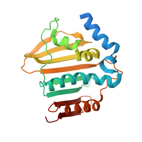
6Y8L, 6Y8N, 6Y8O - PubMed Abstract:
To evaluate the efficacy of two novel compounds against mycobacteria and determine the molecular basis of their action on DNA gyrase using structural and mechanistic approaches. Redx03863 and Redx04739 were tested in antibacterial assays, and also against their target, DNA gyrase, using DNA supercoiling and ATPase assays. X-ray crystallography was used to determine the structure of the gyrase B protein ATPase sub-domain from Mycobacterium smegmatis complexed with the aminocoumarin drug novobiocin, and structures of the same domain from Mycobacterium thermoresistibile complexed with novobiocin, and also with Redx03863. Both compounds, Redx03863 and Redx04739, were active against selected Gram-positive and Gram-negative species, with Redx03863 being the more potent, and Redx04739 showing selectivity against M. smegmatis. Both compounds were potent inhibitors of the supercoiling and ATPase reactions of DNA gyrase, but did not appreciably affect the ATP-independent relaxation reaction. The structure of Redx03863 bound to the gyrase B protein ATPase sub-domain from M. thermoresistibile shows that it binds at a site adjacent to the ATP- and novobiocin-binding sites. We found that most of the mutations that we made in the Redx03863-binding pocket, based on the structure, rendered gyrase inactive. Redx03863 and Redx04739 inhibit gyrase by preventing the binding of ATP. The fact that the Redx03863-binding pocket is distinct from that of novobiocin, coupled with the lack of activity of resistant mutants, suggests that such compounds could have potential to be further exploited as antibiotics.
- Department of Biological Chemistry, John Innes Centre, Norwich Research Park, Norwich NR4 7UH, UK.
Organizational Affiliation: