6Y3P

Crystal structure of the C-terminal domain from K. lactis Pby1, an ATP-grasp enzyme interacting with the mRNA decapping enzyme Dcp2


Experimental Data Snapshot

  • Method: X-RAY DIFFRACTION
  • Resolution: 2.30 Å
  • R-Value Free: 0.240 
  • R-Value Work: 0.188 
  • R-Value Observed: 0.191 

wwPDB Validation   3D Report Full Report


Ligand Structure Quality Assessment 


This is version 1.2 of the entry. See complete history


Literature

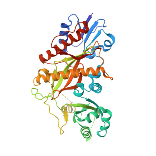
Pby1 is a direct partner of the Dcp2 decapping enzyme.

Charenton, C.Gaudon-Plesse, C.Back, R.Ulryck, N.Cosson, L.Seraphin, B.Graille, M.

(2020) Nucleic Acids Res 48: 6353-6366

  • DOI: https://doi.org/10.1093/nar/gkaa337
  • Primary Citation of Related Structures:  
    6Y3P, 6Y3Z

  • PubMed Abstract: 

    Most eukaryotic mRNAs harbor a characteristic 5' m7GpppN cap that promotes pre-mRNA splicing, mRNA nucleocytoplasmic transport and translation while also protecting mRNAs from exonucleolytic attacks. mRNA caps are eliminated by Dcp2 during mRNA decay, allowing 5'-3' exonucleases to degrade mRNA bodies. However, the Dcp2 decapping enzyme is poorly active on its own and requires binding to stable or transient protein partners to sever the cap of target mRNAs. Here, we analyse the role of one of these partners, the yeast Pby1 factor, which is known to co-localize into P-bodies together with decapping factors. We report that Pby1 uses its C-terminal domain to directly bind to the decapping enzyme. We solved the structure of this Pby1 domain alone and bound to the Dcp1-Dcp2-Edc3 decapping complex. Structure-based mutant analyses reveal that Pby1 binding to the decapping enzyme is required for its recruitment into P-bodies. Moreover, Pby1 binding to the decapping enzyme stimulates growth in conditions in which decapping activation is compromised. Our results point towards a direct connection of Pby1 with decapping and P-body formation, both stemming from its interaction with the Dcp1-Dcp2 holoenzyme.


  • Organizational Affiliation

    Laboratoire de Biologie Structurale de la Cellule (BIOC), CNRS, Ecole polytechnique, IP Paris, 91128 Palaiseau, France.


Macromolecules
Find similar proteins by:  (by identity cutoff)  |  3D Structure
Entity ID: 1
MoleculeChains Sequence LengthOrganismDetailsImage
KLLA0B12012p402Kluyveromyces lactis NRRL Y-1140Mutation(s): 0 
Gene Names: KLLA0_B12012g
UniProt
Find proteins for Q6CVH5 (Kluyveromyces lactis (strain ATCC 8585 / CBS 2359 / DSM 70799 / NBRC 1267 / NRRL Y-1140 / WM37))
Explore Q6CVH5 
Go to UniProtKB:  Q6CVH5
Entity Groups  
Sequence Clusters30% Identity50% Identity70% Identity90% Identity95% Identity100% Identity
UniProt GroupQ6CVH5
Sequence Annotations
Expand
  • Reference Sequence
Experimental Data & Validation

Experimental Data

  • Method: X-RAY DIFFRACTION
  • Resolution: 2.30 Å
  • R-Value Free: 0.240 
  • R-Value Work: 0.188 
  • R-Value Observed: 0.191 
  • Space Group: P 32 2 1
Unit Cell:
Length ( Å )Angle ( ˚ )
a = 95.64α = 90
b = 95.64β = 90
c = 75.84γ = 120
Software Package:
Software NamePurpose
BUSTERrefinement
PDB_EXTRACTdata extraction
XDSdata reduction
XSCALEdata scaling
SHARPphasing

Structure Validation

View Full Validation Report


Ligand Structure Quality Assessment 


Entry History & Funding Information

Deposition Data

  • Released Date: 2020-04-29 
  • Deposition Author(s): Graille, M.

Funding OrganizationLocationGrant Number
ATIP-AvenirFrance--
French National Research AgencyFranceANR-11-BSV800902

Revision History  (Full details and data files)

  • Version 1.0: 2020-04-29
    Type: Initial release
  • Version 1.1: 2020-05-20
    Changes: Database references
  • Version 1.2: 2020-06-24
    Changes: Database references