N-terminal Transmembrane-Helix Epitope Tag for X-ray Crystallography and Electron Microscopy of Small Membrane Proteins.
McIlwain, B.C., Erwin, A.L., Davis, A.R., Ben Koff, B., Chang, L., Bylund, T., Chuang, G.Y., Kwong, P.D., Ohi, M.D., Lai, Y.T., Stockbridge, R.B.(2021) J Mol Biology 433: 166909-166909
- PubMed: 33676924 Search on PubMedSearch on PubMed Central
- DOI: https://doi.org/10.1016/j.jmb.2021.166909
- Primary Citation Related Structures:
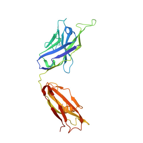
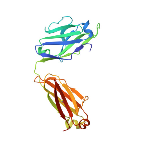
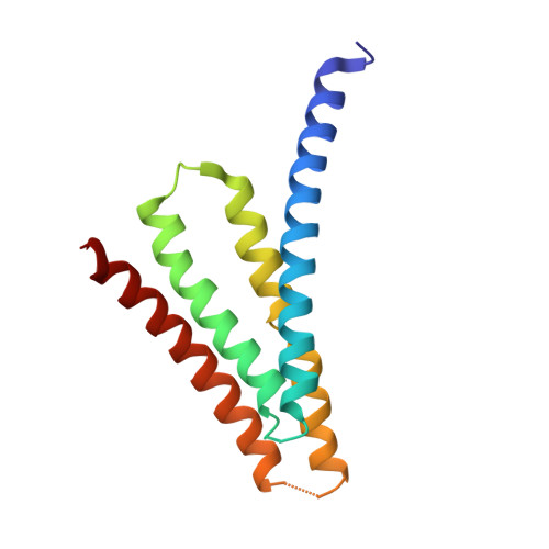
6X58 - PubMed Abstract:
Structural studies of membrane proteins, especially small membrane proteins, are associated with well-known experimental challenges. Complexation with monoclonal antibody fragments is a common strategy to augment such proteins; however, generating antibody fragments that specifically bind a target protein is not trivial. Here we identify a helical epitope, from the membrane-proximal external region (MPER) of the gp41-transmembrane subunit of the HIV envelope protein, that is recognized by several well-characterized antibodies and that can be fused as a contiguous extension of the N-terminal transmembrane helix of a broad range of membrane proteins. To analyze whether this MPER-epitope tag might aid structural studies of small membrane proteins, we determined an X-ray crystal structure of a membrane protein target that does not crystallize without the aid of crystallization chaperones, the Fluc fluoride channel, fused to the MPER epitope and in complex with antibody. We also demonstrate the utility of this approach for single particle electron microscopy with Fluc and two additional small membrane proteins that represent different membrane protein folds, AdiC and GlpF. These studies show that the MPER epitope provides a structurally defined, rigid docking site for antibody fragments that is transferable among diverse membrane proteins and can be engineered without prior structural information. Antibodies that bind to the MPER epitope serve as effective crystallization chaperones and electron microscopy fiducial markers, enabling structural studies of challenging small membrane proteins.
- Department of Molecular, Cellular and Developmental Biology, University of Michigan, Ann Arbor, MI 48109, United States.
Organizational Affiliation: