6X4D

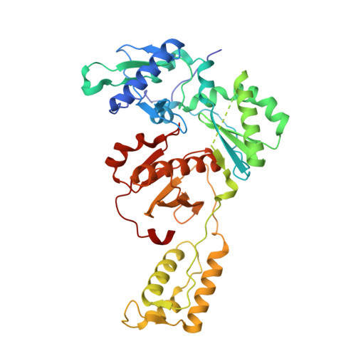
Crystal Structure of HIV-1 Reverse Transcriptase in Complex with 5-(cyclopropylmethyl)-7-(2-(2-(2,4-dioxo-3,4-dihydropyrimidin-1(2H)-yl)ethoxy)phenoxy)-8-methyl-2-naphthonitrile (JLJ678), a Non-nucleoside Inhibitor


Experimental Data Snapshot

  • Method: X-RAY DIFFRACTION
  • Resolution: 2.65 Å
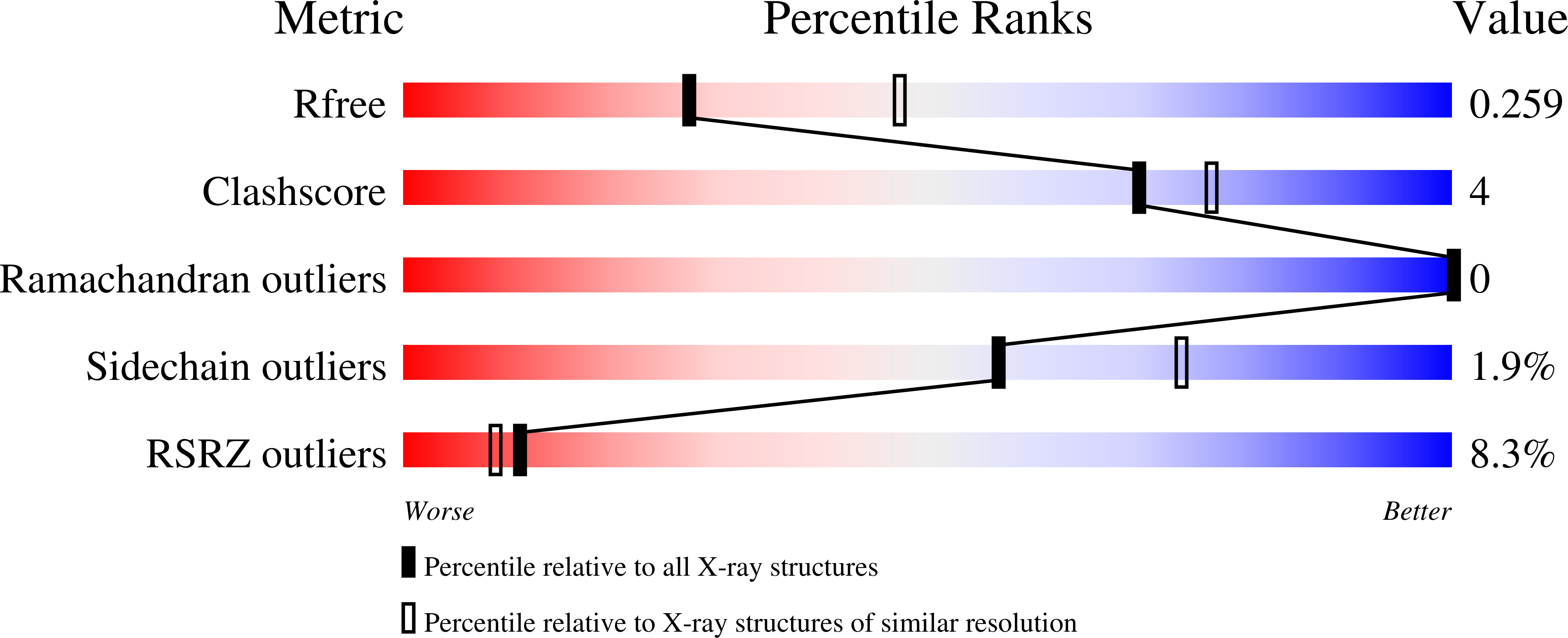
  • R-Value Free: 0.258 
  • R-Value Work: 0.224 
  • R-Value Observed: 0.225 

Starting Model: experimental
View more details

wwPDB Validation   3D Report Full Report


Ligand Structure Quality Assessment 


This is version 1.2 of the entry. See complete history


Literature

Structural investigation of 2-naphthyl phenyl ether inhibitors bound to WT and Y181C reverse transcriptase highlights key features of the NNRTI binding site.

Duong, V.N.Ippolito, J.A.Chan, A.H.Lee, W.G.Spasov, K.A.Jorgensen, W.L.Anderson, K.S.

(2020) Protein Sci 29: 1902-1910

  • DOI: https://doi.org/10.1002/pro.3910
  • Primary Citation of Related Structures:  
    6X47, 6X49, 6X4A, 6X4B, 6X4C, 6X4D, 6X4E, 6X4F

  • PubMed Abstract: 

    Human immunodeficiency virus (HIV)-1 remains as a global health issue that is primarily treated with highly active antiretroviral therapy, a combination of drugs that target the viral life cycle. One class of these drugs are non-nucleoside reverse transcriptase inhibitors (NNRTIs) that target the viral reverse transcriptase (RT). First generation NNRTIs were troubled with poor pharmacological properties and drug resistance, incentivizing the development of improved compounds. One class of developed compounds are the 2-naphthyl phenyl ethers, showing promising efficacy against the Y181C RT mutation. Further biochemical and structural work demonstrated differences in potency against the Y181C mutation and binding mode of the compounds. This work aims to understand the relationship between the binding mode and ability to overcome drug resistance using macromolecular x-ray crystallography. Comparison of 2-naphthyl phenyl ethers bound to Y181C RT reveal that compounds that interact with the invariant W229 are more capable of retaining efficacy against the resistance mutation. Additional modifications to these compounds at the 4-position, computationally designed to compensate for the Y181C mutation, do not demonstrate improved potency. Ultimately, we highlight important considerations for the development of future HIV-1 drugs that are able to combat drug resistance.


  • Organizational Affiliation

    Department of Pharmacology, Yale University School of Medicine, New Haven, Connecticut, USA.


Macromolecules
Find similar proteins by:  (by identity cutoff)  |  3D Structure
Entity ID: 1
MoleculeChains Sequence LengthOrganismDetailsImage
Reverse transcriptase/ribonuclease H557Human immunodeficiency virus type 1 BH10Mutation(s): 3 
Gene Names: gag-pol
EC: 2.7.7.49 (PDB Primary Data), 2.7.7.7 (PDB Primary Data), 3.1.26.13 (PDB Primary Data)
UniProt
Find proteins for P03366 (Human immunodeficiency virus type 1 group M subtype B (isolate BH10))
Explore P03366 
Go to UniProtKB:  P03366
Entity Groups  
Sequence Clusters30% Identity50% Identity70% Identity90% Identity95% Identity100% Identity
UniProt GroupP03366
Sequence Annotations
Expand
  • Reference Sequence
Find similar proteins by:  (by identity cutoff)  |  3D Structure
Entity ID: 2
MoleculeChains Sequence LengthOrganismDetailsImage
p51 RT428Human immunodeficiency virus type 1 BH10Mutation(s): 1 
Gene Names: gag-pol
UniProt
Find proteins for P03366 (Human immunodeficiency virus type 1 group M subtype B (isolate BH10))
Explore P03366 
Go to UniProtKB:  P03366
Entity Groups  
Sequence Clusters30% Identity50% Identity70% Identity90% Identity95% Identity100% Identity
UniProt GroupP03366
Sequence Annotations
Expand
  • Reference Sequence
Small Molecules
Binding Affinity Annotations 
IDSourceBinding Affinity
UO4 BindingDB:  6X4D EC50: min: 900, max: 1200 (nM) from 2 assay(s)
Experimental Data & Validation

Experimental Data

  • Method: X-RAY DIFFRACTION
  • Resolution: 2.65 Å
  • R-Value Free: 0.258 
  • R-Value Work: 0.224 
  • R-Value Observed: 0.225 
  • Space Group: C 1 2 1
Unit Cell:
Length ( Å )Angle ( ˚ )
a = 162.74α = 90
b = 74.27β = 100.02
c = 108.73γ = 90
Software Package:
Software NamePurpose
XSCALEdata scaling
PHENIXrefinement
PDB_EXTRACTdata extraction
XDSdata reduction
PHASERphasing

Structure Validation

View Full Validation Report


Ligand Structure Quality Assessment 


Entry History & Funding Information

Deposition Data


Funding OrganizationLocationGrant Number
National Institutes of Health/National Institute of General Medical Sciences (NIH/NIGMS)United StatesGM49551
National Institutes of Health/National Institute Of Allergy and Infectious Diseases (NIH/NIAID)United StatesAI155072

Revision History  (Full details and data files)

  • Version 1.0: 2020-07-22
    Type: Initial release
  • Version 1.1: 2020-12-16
    Changes: Database references
  • Version 1.2: 2023-10-18
    Changes: Data collection, Database references, Refinement description