6WYE

Crystal structure of Neisseria gonorrhoeae serine acetyltransferase (CysE)


Experimental Data Snapshot

  • Method: X-RAY DIFFRACTION
  • Resolution: 2.01 Å
  • R-Value Free: 0.222 
  • R-Value Work: 0.183 
  • R-Value Observed: 0.185 

wwPDB Validation   3D Report Full Report


This is version 1.3 of the entry. See complete history


Literature

Serine acetyltransferase from Neisseria gonorrhoeae; structural and biochemical basis of inhibition.

Oldham, K.E.A.Prentice, E.J.Summers, E.L.Hicks, J.L.

(2022) Biochem J 479: 57-74

  • DOI: https://doi.org/10.1042/BCJ20210564
  • Primary Citation of Related Structures:  
    6WYE, 7RA4

  • PubMed Abstract: 

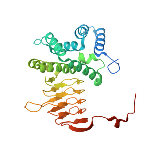
    Serine acetyltransferase (SAT) catalyzes the first step in the two-step pathway to synthesize l-cysteine in bacteria and plants. SAT synthesizes O-acetylserine from substrates l-serine and acetyl coenzyme A and is a key enzyme for regulating cellular cysteine levels by feedback inhibition of l-cysteine, and its involvement in the cysteine synthase complex. We have performed extensive structural and kinetic characterization of the SAT enzyme from the antibiotic-resistant pathogen Neisseria gonorrhoeae. Using X-ray crystallography, we have solved the structures of NgSAT with the non-natural ligand, l-malate (present in the crystallization screen) to 2.01 Å and with the natural substrate l-serine (2.80 Å) bound. Both structures are hexamers, with each monomer displaying the characteristic left-handed parallel β-helix domain of the acyltransferase superfamily of enzymes. Each structure displays both extended and closed conformations of the C-terminal tail. l-malate bound in the active site results in an interesting mix of open and closed active site conformations, exhibiting a structural change mimicking the conformation of cysteine (inhibitor) bound structures from other organisms. Kinetic characterization shows competitive inhibition of l-cysteine with substrates l-serine and acetyl coenzyme A. The SAT reaction represents a key point for the regulation of cysteine biosynthesis and controlling cellular sulfur due to feedback inhibition by l-cysteine and formation of the cysteine synthase complex. Data presented here provide the structural and mechanistic basis for inhibitor design and given this enzyme is not present in humans could be explored to combat the rise of extensively antimicrobial resistant N. gonorrhoeae.


  • Organizational Affiliation

    Te Aka Mātuatua School of Science, University of Waikato, Hamilton, New Zealand.


Macromolecules
Find similar proteins by:  (by identity cutoff)  |  3D Structure
Entity ID: 1
MoleculeChains Sequence LengthOrganismDetailsImage
Serine acetyltransferase
A, B, C, D, E
A, B, C, D, E, F
293Neisseria gonorrhoeaeMutation(s): 0 
Gene Names: cysEE8M63_06755F0T10_08440F0T11_08395F0T12_08425NCTC13805_01597WHOF_01434WHOF_01635
EC: 2.3.1.30
UniProt
Find proteins for Q5F6X0 (Neisseria gonorrhoeae (strain ATCC 700825 / FA 1090))
Explore Q5F6X0 
Go to UniProtKB:  Q5F6X0
Entity Groups  
Sequence Clusters30% Identity50% Identity70% Identity90% Identity95% Identity100% Identity
UniProt GroupQ5F6X0
Sequence Annotations
Expand
  • Reference Sequence
Small Molecules
Ligands 2 Unique
IDChains Name / Formula / InChI Key2D Diagram3D Interactions
LMR
Query on LMR

Download Ideal Coordinates CCD File 
H [auth A]
I [auth A]
K [auth B]
N [auth D]
P [auth E]
H [auth A],
I [auth A],
K [auth B],
N [auth D],
P [auth E],
R [auth F]
(2S)-2-hydroxybutanedioic acid
C4 H6 O5
BJEPYKJPYRNKOW-REOHCLBHSA-N
NA
Query on NA

Download Ideal Coordinates CCD File 
G [auth A]
J [auth B]
L [auth C]
M [auth D]
O [auth E]
G [auth A],
J [auth B],
L [auth C],
M [auth D],
O [auth E],
Q [auth F]
SODIUM ION
Na
FKNQFGJONOIPTF-UHFFFAOYSA-N
Experimental Data & Validation

Experimental Data

  • Method: X-RAY DIFFRACTION
  • Resolution: 2.01 Å
  • R-Value Free: 0.222 
  • R-Value Work: 0.183 
  • R-Value Observed: 0.185 
  • Space Group: P 1 2 1
Unit Cell:
Length ( Å )Angle ( ˚ )
a = 77.897α = 90
b = 93.867β = 91.29
c = 102.019γ = 90
Software Package:
Software NamePurpose
PHENIXrefinement
XDSdata reduction
XDSdata scaling
PHASERphasing

Structure Validation

View Full Validation Report


Entry History & Funding Information

Deposition Data


Funding OrganizationLocationGrant Number
Health Research Council (HRC)New Zealand19/602

Revision History  (Full details and data files)

  • Version 1.0: 2021-06-09
    Type: Initial release
  • Version 1.1: 2021-12-22
    Changes: Database references
  • Version 1.2: 2022-01-19
    Changes: Database references
  • Version 1.3: 2023-10-18
    Changes: Data collection, Refinement description